. 2011 Nov 18;415(2):319-24.
doi: 10.1016/j.bbrc.2011.10.052. Epub 2011 Oct 18.

Muscarinic receptor agonists stimulate matrix metalloproteinase 1-dependent invasion of human colon cancer cells

Affiliations

Muscarinic receptor agonists stimulate matrix metalloproteinase 1-dependent invasion of human colon cancer cells

Jean-Pierre Raufman et al. Biochem Biophys Res Commun. .

Abstract

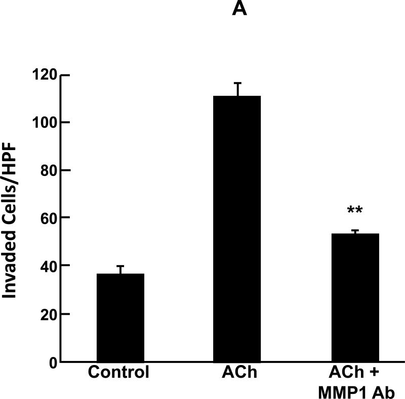
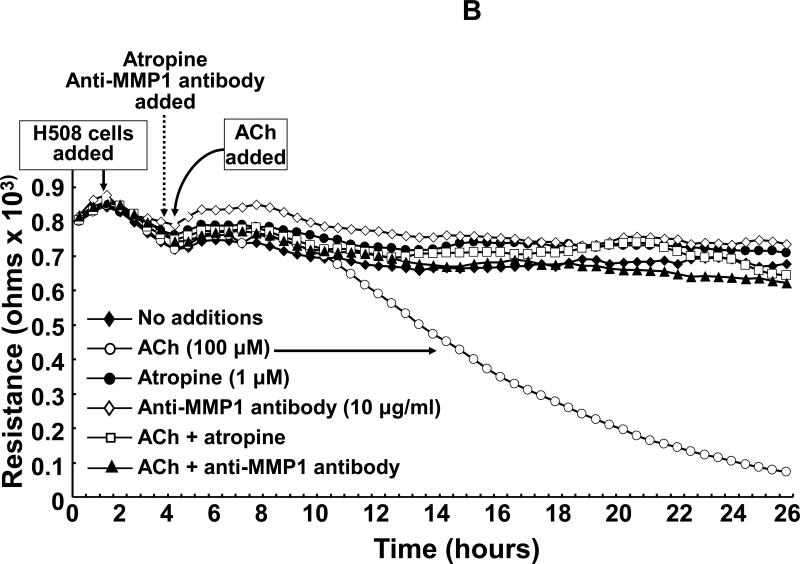
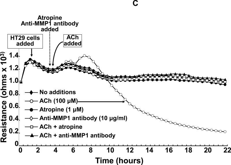
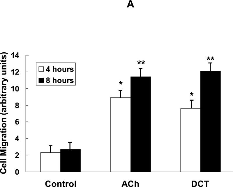
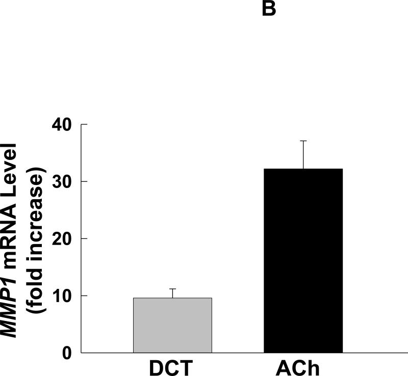
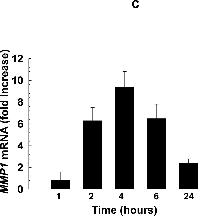
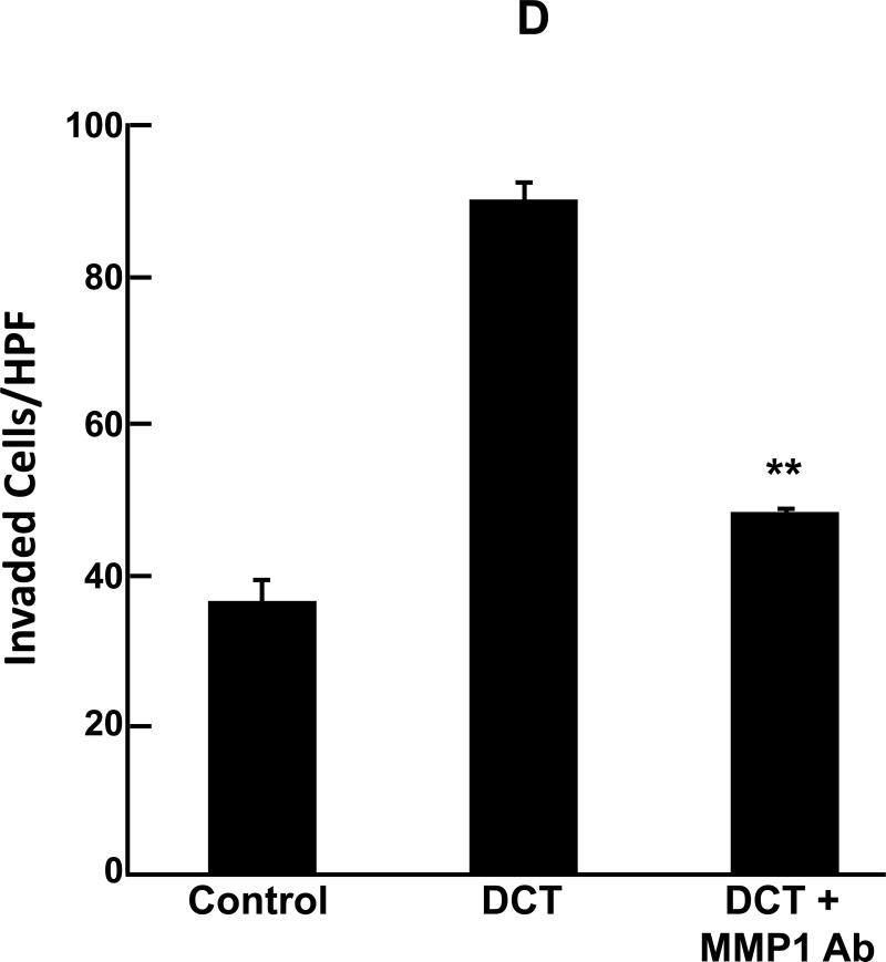
Mammalian matrix metalloproteinases (MMPs) which degrade extracellular matrix facilitate colon cancer cell invasion into the bloodstream and extra-colonic tissues; in particular, MMP1 expression correlates strongly with advanced colon cancer stage, hematogenous metastasis and poor prognosis. Likewise, muscarinic receptor signaling plays an important role in colon cancer; muscarinic receptors are over-expressed in colon cancer compared to normal colon epithelial cells. Muscarinic receptor activation stimulates proliferation, migration and invasion of human colon cancer cells. In mouse intestinal neoplasia models genetic ablation of muscarinic receptors attenuates carcinogenesis. In the present work, we sought to link these observations by showing that MMP1 expression and activation plays a mechanistic role in muscarinic receptor agonist-induced colon cancer cell invasion. We show that acetylcholine, which robustly increases MMP1 expression, stimulates invasion of HT29 and H508 human colon cancer cells into human umbilical vein endothelial cell monolayers - this was abolished by pre-incubation with atropine, a non-selective muscarinic receptor inhibitor, and by pre-incubation with anti-MMP1 neutralizing antibody. Similar results were obtained using a Matrigel chamber assay and deoxycholyltaurine (DCT), an amidated dihydroxy bile acid associated with colon neoplasia in animal models and humans, and previously shown to interact functionally with muscarinic receptors. DCT treatment of human colon cancer cells resulted in time-dependent, 10-fold increased MMP1 expression, and DCT-induced cell invasion was also blocked by pre-treatment with anti-MMP1 antibody. This study contributes to understanding mechanisms underlying muscarinic receptor agonist-induced promotion of colon cancer and, more importantly, indicates that blocking MMP1 expression and activation has therapeutic promise to stop or retard colon cancer invasion and dissemination.


Figures

Fig. 1
Fig. 1
Acetylcholine (ACh) stimulates MMP1-dependent human colon cancer cell invasion. A: HT29 colon cancer cells were incubated for 24 h with ACh (100 μM), alone or in cells pre-incubated for 30 min with anti-MMP1 antibody (10 μg/ml), and invasion was measured using BD Biocoat Invasion Chambers with Matrigel inserts. Values represent mean ± SE (3 separate experiments). Anti-MMP1 antibody alone did not alter basal HT29 cell invasion (not shown). HPF, high power field. **, P < 0.01, Student's paired t-test. B: Electrical cell impedance sensing (ECIS) shows ACh-induced H508 colon cancer cell invasion of HUVEC monolayers. At the time shown, ACh (100 μM) was added to H508 cells, with or without pre-incubation for 30 min with atropine (1 μM) or anti-MMP1 antibody (10 μg/ml), and invasion into the HUVEC monolayer was determined by measuring trans-HUVEC monolayer resistance. ACh-induced H508 cell invasion into the HUVEC monolayer was blocked by both atropine and anti-MMP1 antibody. In additional control experiments, the agents tested had no effect on HUVEC monolayer resistance in the absence of added H508 cells (not shown). C: ECIS experiments were conducted with HT29 cells instead of H508 cells using the same experimental approach and controls described for Fig. 1B. For both H508 and HT29 cells, resistance (ohms) of the HUVEC monolayer over the course of the experiments (mean of duplicates at each time point) was representative of three separate experiments.
Fig. 1
Fig. 1
Acetylcholine (ACh) stimulates MMP1-dependent human colon cancer cell invasion. A: HT29 colon cancer cells were incubated for 24 h with ACh (100 μM), alone or in cells pre-incubated for 30 min with anti-MMP1 antibody (10 μg/ml), and invasion was measured using BD Biocoat Invasion Chambers with Matrigel inserts. Values represent mean ± SE (3 separate experiments). Anti-MMP1 antibody alone did not alter basal HT29 cell invasion (not shown). HPF, high power field. **, P < 0.01, Student's paired t-test. B: Electrical cell impedance sensing (ECIS) shows ACh-induced H508 colon cancer cell invasion of HUVEC monolayers. At the time shown, ACh (100 μM) was added to H508 cells, with or without pre-incubation for 30 min with atropine (1 μM) or anti-MMP1 antibody (10 μg/ml), and invasion into the HUVEC monolayer was determined by measuring trans-HUVEC monolayer resistance. ACh-induced H508 cell invasion into the HUVEC monolayer was blocked by both atropine and anti-MMP1 antibody. In additional control experiments, the agents tested had no effect on HUVEC monolayer resistance in the absence of added H508 cells (not shown). C: ECIS experiments were conducted with HT29 cells instead of H508 cells using the same experimental approach and controls described for Fig. 1B. For both H508 and HT29 cells, resistance (ohms) of the HUVEC monolayer over the course of the experiments (mean of duplicates at each time point) was representative of three separate experiments.
Fig. 2
Fig. 2
Deoxycholyltaurine (DCT), a dihydroxy bile acid that interacts functionally with muscarinic receptors, stimulates colon cancer cell migration and MMP1-dependent invasion. A: DCT (100 μM) stimulates H508 cell migration in a scratch-wound assay. Cells were plated at confluence before linear wounding. Photomicrographs were taken before adding test agents and at 4 and 8 hours after adding ACh and DCT. Values for ACh and DCT were greater than vehicle (control) at both time points (N > 3 expts; *, P < 0.05; **, P < 0.01, ANOVA with Tukey's test). B: Comparison of DCT- and ACh-induced MMP1 mRNA expression in human colon cancer cells. qPCR was performed using cDNA synthesized from total RNA prepared from H508 cells after incubation with MR agonists. MMP1 mRNA levels were measured after 4 h incubation with maximal concentrations of DCT (50 μM) and ACh (100 μM). Mean ± SE from at least 3 experiments. C: Time course for DCT-induced MMP1 expression. qPCR was performed using cDNA synthesized from total RNA prepared from H508 cells incubated for the times indicated with DCT (50 μM). Mean ± SE from at least 3 experiments. D: HT29 colon cancer cells were incubated for 24 h with DCT (100 μM), alone or in cells pre-incubated for 30 min with anti-MMP1 antibody (10 μg/ml). Cell invasion was measured using BD Biocoat Invasion Chambers with Matrigel inserts. Values represent mean ± SE (3 separate experiments). Anti-MMP1 antibody alone did not alter basal HT29 cell invasion (not shown). HPF, high power field. **, P < 0.01, Student's paired t-test.
Fig. 2
Fig. 2
Deoxycholyltaurine (DCT), a dihydroxy bile acid that interacts functionally with muscarinic receptors, stimulates colon cancer cell migration and MMP1-dependent invasion. A: DCT (100 μM) stimulates H508 cell migration in a scratch-wound assay. Cells were plated at confluence before linear wounding. Photomicrographs were taken before adding test agents and at 4 and 8 hours after adding ACh and DCT. Values for ACh and DCT were greater than vehicle (control) at both time points (N > 3 expts; *, P < 0.05; **, P < 0.01, ANOVA with Tukey's test). B: Comparison of DCT- and ACh-induced MMP1 mRNA expression in human colon cancer cells. qPCR was performed using cDNA synthesized from total RNA prepared from H508 cells after incubation with MR agonists. MMP1 mRNA levels were measured after 4 h incubation with maximal concentrations of DCT (50 μM) and ACh (100 μM). Mean ± SE from at least 3 experiments. C: Time course for DCT-induced MMP1 expression. qPCR was performed using cDNA synthesized from total RNA prepared from H508 cells incubated for the times indicated with DCT (50 μM). Mean ± SE from at least 3 experiments. D: HT29 colon cancer cells were incubated for 24 h with DCT (100 μM), alone or in cells pre-incubated for 30 min with anti-MMP1 antibody (10 μg/ml). Cell invasion was measured using BD Biocoat Invasion Chambers with Matrigel inserts. Values represent mean ± SE (3 separate experiments). Anti-MMP1 antibody alone did not alter basal HT29 cell invasion (not shown). HPF, high power field. **, P < 0.01, Student's paired t-test.

Similar articles

Cited by

References

    1. Westermarck J, Kahari VM. Regulation of matrix metalloproteinase expression in tumor invasion. FASEB J. 1999;13:781–792. - PubMed
    1. Zucker S, Vacirca J. Role of matrix metalloproteinases (MMPs) in colorectal cancer. Cancer Metastasis Rev. 2004;23:101–117. - PubMed
    1. Murray GI, Duncan ME, O'Neil P, Melvin WT, Fothergill JE. Matrix metalloproteinase-1 is associated with poor prognosis in colorectal cancer. Nat Med. 1996;2:461–462. - PubMed
    1. Baker EA, Bergin FG, Leaper DJ. Matrix metalloproteinases, their tissue inhibitors and colorectal cancer staging. Br J Surg. 2000;87:1215–1221. - PubMed
    1. Sunami E, Tsuno N, Osada T, Saito S, Kitayama J, Tomozawa S, Tsuruo T, Shibata Y, Muto T, Nagawa H. MMP-1 is a prognostic marker for hematogenous metastasis of colorectal cancer. Oncologist. 2000;5:108–114. - PubMed

Publication types

MeSH terms

LinkOut - more resources