. 2012 Jan 2;287(1):91-104.
doi: 10.1074/jbc.M111.296020. Epub 2011 Oct 25.

Modulation of L-α-lysophosphatidylinositol/GPR55 mitogen-activated protein kinase (MAPK) signaling by cannabinoids

Affiliations

Modulation of L-α-lysophosphatidylinositol/GPR55 mitogen-activated protein kinase (MAPK) signaling by cannabinoids

Sharon Anavi-Goffer et al. J Biol Chem. .

Abstract

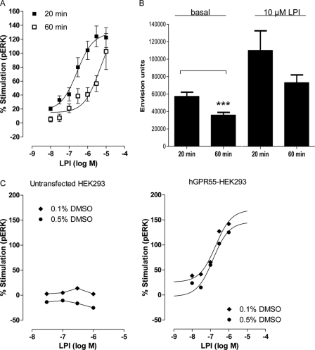
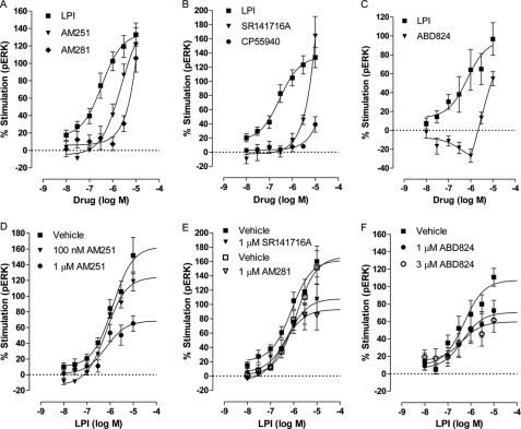
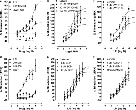
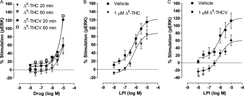
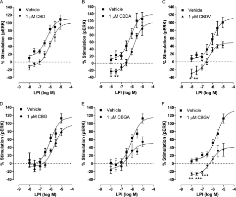
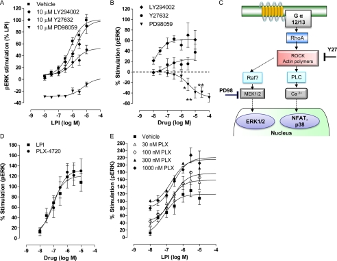
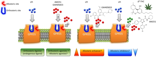
GPR55 is activated by l-α-lysophosphatidylinositol (LPI) but also by certain cannabinoids. In this study, we investigated the GPR55 pharmacology of various cannabinoids, including analogues of the CB1 receptor antagonist Rimonabant®, CB2 receptor agonists, and Cannabis sativa constituents. To test ERK1/2 phosphorylation, a primary downstream signaling pathway that conveys LPI-induced activation of GPR55, a high throughput system, was established using the AlphaScreen® SureFire® assay. Here, we show that CB1 receptor antagonists can act both as agonists alone and as inhibitors of LPI signaling under the same assay conditions. This study clarifies the controversy surrounding the GPR55-mediated actions of SR141716A; some reports indicate the compound to be an agonist and some report antagonism. In contrast, we report that the CB2 ligand GW405833 behaves as a partial agonist of GPR55 alone and enhances LPI signaling. GPR55 has been implicated in pain transmission, and thus our results suggest that this receptor may be responsible for some of the antinociceptive actions of certain CB2 receptor ligands. The phytocannabinoids Δ9-tetrahydrocannabivarin, cannabidivarin, and cannabigerovarin are also potent inhibitors of LPI. These Cannabis sativa constituents may represent novel therapeutics targeting GPR55.


Figures

FIGURE 1.
FIGURE 1.
Detection of LPI-induced ERK1/2 phosphorylation in hGPR55-HEK293 cells using the AlphaScreen® SureFire® ERK assay. A, mean log concentration-response curves for the effect of LPI on ERK1/2 phosphorylation in hGPR55-HEK293 cells after 20 min (n = 4 each in triplicate) or 60 min (n = 3 each 2–3 repeats) of incubation at 37 °C. Increasing the incubation time significantly (p < 0.05) reduced the potency of LPI-induced ERK1/2 phosphorylation but did not affect LPI-induced maximal stimulation (Emax) of the GPR55 receptor. B, compared with a 20-min incubation time at 37 °C, a 60-min incubation significantly (p < 0.05) reduced the basal level of ERK1/2 phosphorylation (n = 4, each in 12 repeats). Raw data of AlphaScreen® SureFire® ERK assay are presented as Envision units, Student's t test, ***, p < 0.05. C, no significant differences were observed in basal levels of phosphorylated ERK1/2 in untransfected HEK293 and hGPR55-HEK293 incubated for 20 min in 0.1 or 0.5% DMSO. C, effect of DMSO on LPI-induced ERK1/2 phosphorylation was assessed at 0.1 or 0.5% DMSO in either hGPR55-HEK293 cells or untransfected HEK293 cells. In untransfected HEK293 cells, no stimulation of ERK1/2 phosphorylation was detected at any concentration of LPI at any concentration of DMSO (untransfected and hGPR55-HEK293 cells were seeded on the same plate). Each symbol represents the mean percentage change in bound phosphorylated ERK1/2 protein.
FIGURE 2.
FIGURE 2.
Structural analogues of SR141716A-induced ERK1/2 phosphorylation in hGPR55-HEK293 cells. A, mean log concentration-response curves for percent stimulation of ERK1/2 phosphorylation by LPI (n = 4), AM251 (n = 3), or AM281 (n = 3) after a 20 min stimulation at 37 °C. B, mean log concentration-response curves of LPI (n = 4), SR141716A (n = 3), or CP55940 (n = 3), a nonselective cannabinoid CB1/CB2 agonist. C, mean log concentration-response curves of LPI or ABD824, an AM251 analog in which iodine was substituted with a bromine (n = 3, each in duplicate). The effect of SR141716A analogues on LPI induced ERK1/2 phosphorylation in hGPR55-HEK293 cells. D, LPI (n = 4) in the presence or absence of 100 nm AM251 (n = 3) or 1 μm AM251 (n = 4). E, effect of LPI in the presence or absence of 1 μm SR141716A (n = 4) or 1 μm AM281 (n = 3). F, effect of LPI in the presence or absence of 1 μm and 3 μm ABD824 (n = 4, each in duplicate). Each symbol represents the mean percentage change in bound phosphorylated ERK1/2 ± S.E. over the basal level, and each independent experiment was performed in triplicate unless stated otherwise.
FIGURE 3.
FIGURE 3.
Effect of CB2 receptor agonists on ERK1/2 phosphorylation in hGPR55-HEK293 cells. A, mean log concentration-response curves for percent stimulation of ERK1/2 phosphorylation by LPI (n = 7), GW405833 (n = 4), or JWH-133 (n = 3) after 20 min of stimulation at 37 °C. JWH-133 significantly reduced basal pERK levels, *, p < 0.05; **, p < 0.01, one-sample t test. B, GW405833 at 10, 30, and 100 nm enhanced the LPI-induced ERK1/2 phosphorylation (n = 4). C, JWH-133 at 1 and 3 μm inhibited the LPI-induced ERK1/2 phosphorylation (n = 4). D, percent stimulation of ERK1/2 phosphorylation by LPI (n = 3), AM1241 (n = 4), or HU-308 (n = 4) and (E)-β-caryophyllene (BCP) (n = 3) after 20 min of stimulation at 37 °C. E, (E)-β-caryophyllene at 1, 3, and 10 μm (n = 3) did not alter the LPI-induced ERK1/2 phosphorylation nor did AM1241 (F, n = 3). Each symbol represents the mean percentage change in bound phosphorylated ERK1/2 ± S.E. over the basal level, and each independent experiment was performed in duplicate.
FIGURE 4.
FIGURE 4.
Effect of Δ9-THC and Δ9-THCV on ERK1/2 phosphorylation in hGPR55-HEK293 cells. Mean log concentration-response curves for ERK1/2 phosphorylation after 20 or 60 min of stimulation at 37 °C with Δ9-THC (n = 3) or Δ9-THCV (n = 4) (A). B shows the effect of LPI in the presence or absence of 1 μm Δ9-THC after 20 min of stimulation at 37 °C (n = 4). C shows the effect of LPI in the presence or absence of 1 μm Δ9-THCV after 20 min of stimulation at 37 °C (n = 3). Each symbol represents the mean percentage change in bound phosphorylated ERK1/2 ± S.E. over the basal level, and each independent experiment was performed in triplicate.
FIGURE 5.
FIGURE 5.
Effect of CBD and CBDA and CBDV on ERK1/2 phosphorylation in hGPR55-HEK293 cells. Mean log concentration-response curves for ERK1/2 phosphorylation after 20 min of stimulation at 37 °C. The effect of LPI in the presence or absence is shown as follows: 1 μm CBD (n = 5) (A); 1 μm CBDA (n = 3) (B); 1 μm CBDV (n = 3) (C); 1 μm CBG (n = 4) (D); 1 μm CBGA (n = 4) (E); 1 μm CBGV (n = 3) (F). CBDV and CGV pronouncedly inhibited LPI compared with their analogues. Each symbol represents the mean percentage change in bound phosphorylated ERK1/2 ± S.E. over the basal level (n = 3). CBDA, CBDV, and CBGV significantly reduced basal pERK levels, *, p < 0.05; **, p < 0.01; ***, p < 0.001 one-sample t test.
FIGURE 6.
FIGURE 6.
Effect of kinase inhibitors. A, LPI-induced stimulation of ERK1/2 phosphorylation was attenuated by 10 μm PD98059, a MEK1 inhibitor, and inhibited by 10 μm Y27632, a Rho-associated protein kinase inhibitor (n = 5 each in duplicate), but not by 10 μm LY294002, a phosphatidylinositol 3-kinase inhibitor (n = 3 each in duplicate). B, effect of each inhibitor alone on phosphorylated ERK1/2 level (n = 4 for PD98059; n = 7 for Y27632; and n = 3 for LY294002, each in duplicate). PD98059 significantly reduced basal pERK levels; *, p < 0.05; **, p < 0.01, one-sample t test. C, diagram representing the putative GPR55/LPI signaling cascade. D, selective B-Raf inhibitor, PLX-4720, stimulated hGPR55-HEK293-expressing cells in a similar manner to LPI (n = 3 each in duplicate). E, increasing concentrations of PLX-4720 significantly increased the efficacy of LPI-induced response (100 nm, p < 0.05; 300 nm, p < 0.01; 1000 nm, p < 0.01) and the bottom of the curve (300 nm, p < 0.05), (n = 3 each in duplicate).
FIGURE 7.
FIGURE 7.
Pharmacological mechanisms for the modulation of GPR55. LPI is suggested to primarily bind the GPR55 orthosteric binding site. GPR55 may also contain an allosteric binding site. These observations raise at least two possibilities as follows. Left, one possibility is that certain arylpyrazole ligands actually represent bitopic ligands of GPR55. These ligands may have the capacity to modulate both the orthosteric (agonists) and the allosteric site through different pharmacophores. The second possibility is that AM251 and certain arylpyrazole analogues are only allosteric ligands that are ago-allosteric alone. Right, in the presence of LPI, arylpyrazoles can also behave as allosteric inhibitors and GW405833 as an allosteric enhancer. In addition, a number of Cannabis sativa constituents appear to inhibit ERK1/2 phosphorylation in an allosteric manner.

References

    1. Staton P. C., Hatcher J. P., Walker D. J., Morrison A. D., Shapland E. M., Hughes J. P., Chong E., Mander P. K., Green P. J., Billinton A., Fulleylove M., Lancaster H. C., Smith J. C., Bailey L. T., Wise A., Brown A. J., Richardson J. C., Chessell I. P. (2008) Pain 139, 225–236 - PubMed
    1. Whyte L. S., Ryberg E., Sims N. A., Ridge S. A., Mackie K., Greasley P. J., Ross R. A., Rogers M. J. (2009) Proc. Natl. Acad. Sci. U.S.A. 106, 16511–16516 - PMC - PubMed
    1. Ford L. A., Roelofs A. J., Anavi-Goffer S., Mowat L., Simpson D. G., Irving A. J., Rogers M. J., Rajnicek A. M., Ross R. A. (2010) Br. J. Pharmacol. 160, 762–771 - PMC - PubMed
    1. Henstridge C. M., Balenga N. A., Ford L. A., Ross R. A., Waldhoer M., Irving A. J. (2009) FASEB J. 23, 183–193 - PubMed
    1. Kapur A., Zhao P., Sharir H., Bai Y., Caron M. G., Barak L. S., Abood M. E. (2009) J. Biol. Chem. 284, 29817–29827 - PMC - PubMed

Publication types

MeSH terms

LinkOut - more resources