. 2011 Dec 30;286(52):44691-701.
doi: 10.1074/jbc.M111.303917. Epub 2011 Oct 25.

The intracellular amino terminus plays a dominant role in desensitization of ATP-gated P2X receptor ion channels

Affiliations

The intracellular amino terminus plays a dominant role in desensitization of ATP-gated P2X receptor ion channels

Rebecca C Allsopp et al. J Biol Chem. .

Abstract

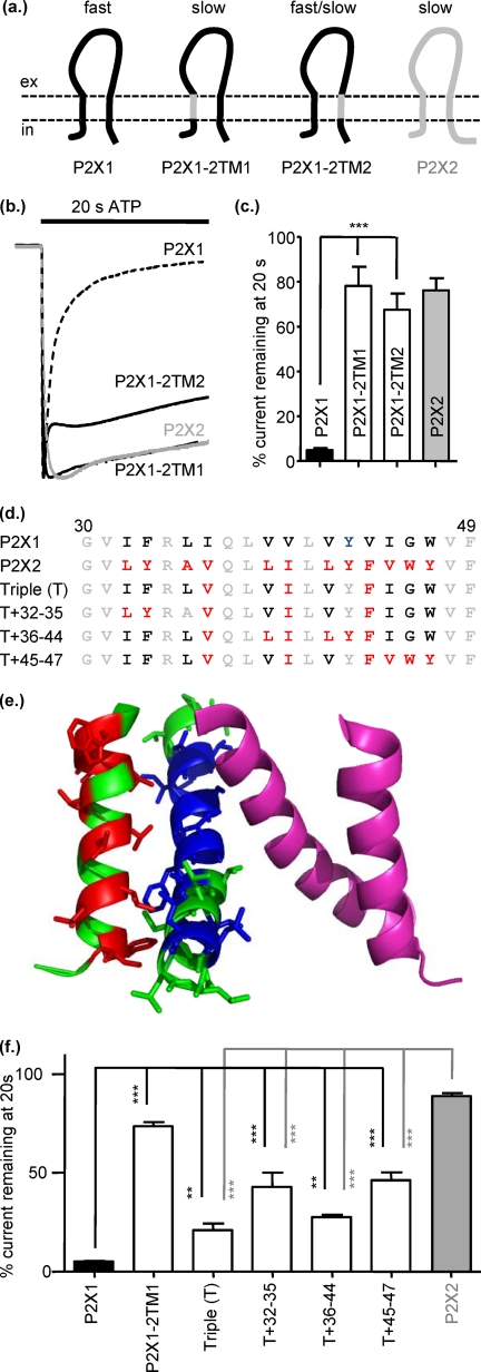
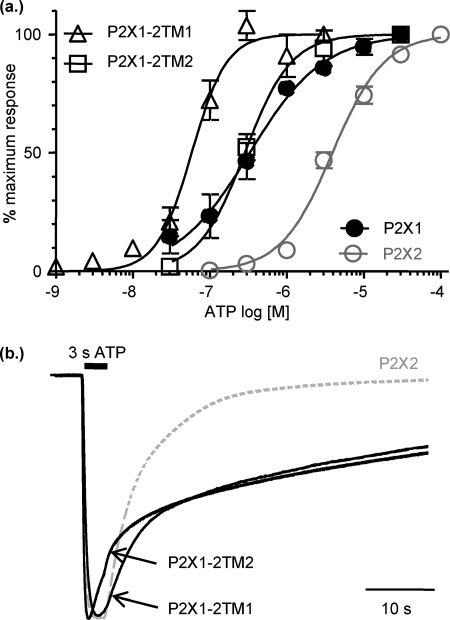
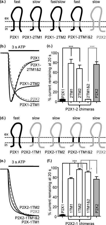
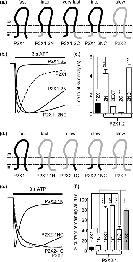
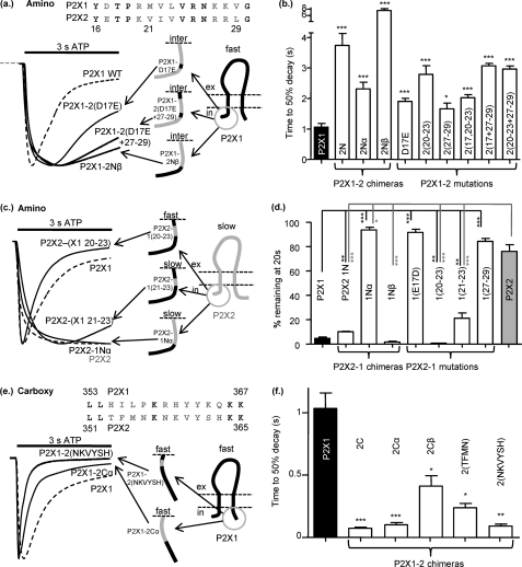
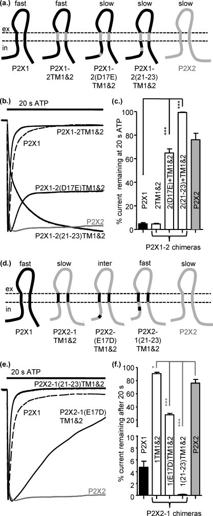
P2X receptors show marked variations in the time-course of response to ATP application from rapidly desensitizing P2X1 receptors to relatively sustained P2X2 receptors. In this study we have used chimeras between human P2X1 and P2X2 receptors in combination with mutagenesis to address the contribution of the extracellular ligand binding loop, the transmembrane channel, and the intracellular regions to receptor time-course. Swapping either the extracellular loop or both transmembrane domains (TM1 and -2) between the P2X1 and P2X2 receptors had no effect on the time-course of ATP currents in the recipient receptor. These results suggest that the agonist binding and channel-forming portions of the receptor do not play a major role in the control of the time-course. In contrast replacing the amino terminus of the P2X1 receptor with that from the non-desensitizing P2X2 receptor (P2X1-2N) slowed desensitization, and the mirror chimera induced rapid desensitization in the P2X2-1N chimera. These reciprocal effects on time-course can be replicated by changing four variant amino acids just before the first transmembrane (TM1) segment. These pre-TM1 residues also had a dominant effect on chimeras where both TMs had been transferred; mutating the variant amino acids 21-23 to those found in the P2X2 receptor removed desensitization from the P2X1-2TM1/-2 chimera, and the reciprocal mutants induced rapid desensitization in the non-desensitizing P2X2-1TM1/-2 chimera. These results suggest that the intracellular amino terminus, in particular the region just before TM1, plays a dominant role in the regulation of the time-course of ATP evoked P2X receptor currents.


Figures

FIGURE 1.
FIGURE 1.
Replacing either of the transmembrane domains of P2X1 with those from P2X2 reduced desensitization. a, schematic shows P2X1 (black) and P2X2 (gray) receptors and the chimeras P2X1-2TM1 and P2X1-2TM2. b, representative currents mediated by application of 100 μm ATP to P2X1 (dotted line), P2X2 (gray line), and chimeric receptors expressed in Xenopus oocytes. c, histogram summary shows the percentage of current remaining at the end of a 20-s ATP application. d, shown is an amino acid sequence lineup of P2X1 and P2X2 TM1. Conserved amino acids are shown in gray, variant amino acids are in black, and mutants of non-conserved amino acids are shown in red. e, a P2X1 receptor homology model shows the TM regions for two subunits (green and magenta); the green subunit non-conserved amino acids in TM1 are shown in red and in blue for TM2. f, shown is a summary of the percentage current remaining at 20s to100 μm ATP application. *, p < 0.05; **, p < 0.01; ***, p < 0.001 (n = 3–10).
FIGURE 2.
FIGURE 2.
Prolongation of current deactivation after ATP washout for P2X1-2TM chimeras. a, shown are concentration responses to ATP for P2X2 (gray line) and chimeric P2X1-2TM1 and P2X1-2TM2 receptors (n = 3–5). b, shown are time-course of currents evoked by a 3-s application of an EC75 concentration of ATP (P2X2 10 μm, P2X1-2TM1 0.1 μm, P2X1-2TM2 1 μm). ATP was applied for 3 s as shown by the black bar. Traces are normalized to the peak current to highlight the prolonged deactivation for the chimeric receptors on washout of ATP.
FIGURE 3.
FIGURE 3.
Swapping both TMs between P2X1 and P2X2 receptors has no effect on the time-course of response. a, shown is a schematic representation of P2X1 (black), P2X2 (gray), and chimeric receptors. b, representative currents mediated by a 3-s application of 100 μm ATP to P2X1 (black dotted line), P2X2 (gray dotted line), and chimeric P2X receptors expressed in Xenopus oocytes. c, a histogram summary shows the percentage of current remaining at the end of a 20-s ATP application. d, shown is a schematic of the reciprocal chimeras on a P2X2 receptor background. e, representative currents are mediated by a 3-s application of 100 μm ATP. f, a histogram summary show the percentage of current remaining at the end of a 20-s 100 μm ATP application. *, p < 0.05; ***, p < 0.001 (n = 3–10).
FIGURE 4.
FIGURE 4.
Contribution of the intracellular regions to regulation of P2X receptor time-course. a, shown is a schematic representation of P2X receptors and chimeras. inter, intermediate. b, representative currents were mediated by application of 100 μm ATP to chimeric receptors expressed in Xenopus oocytes. c, a histogram summary shows the time to 50% current decay during the continued presence of ATP. d, shown is a schematic representation of P2X receptors and chimeras. e, representative currents were mediated by application of 100 μm ATP to chimeric receptors expressed in Xenopus oocytes. c, a histogram summary shows the percentage of current remaining at the end of a 20-s 100 μm ATP application. **, p < 0.01; ***, p < 0.001 (n = 5–17).
FIGURE 5.
FIGURE 5.
Regions of the intracellular amino and carboxyl termini involved in regulating time-course. a, shown is a schematic representation of chimeras and representative currents (to 100 μm ATP) of P2X1-2N, P2X1-2Nβ, and P2X1-2 mutations. Mutations are based on the non-conserved amino acid residues within the Nβ region between P2X1 and P2X2 (shown in gray in the amino acid lineup, P2X1 receptor numbering). b, a histogram summary shows the time to 50% decay during continued ATP application. c, shown is a schematic representation and representative currents (to 100 μm ATP) of the reciprocal set of chimeras and P2X2-1 mutations. d, a histogram summary shows the percentage of current remaining at the end of a 20-s 100 μm ATP application. e, a schematic representation and representative currents (to 100 μm ATP) of P2X1-2Cα and P2X1-2 mutations of non-conserved amino acids within this region (non-conserved amino acids between P2X1 and P2X2 are shown in gray on the sequence lineup). f, a histogram summary shows the time to 50% decay during continued ATP application. *, p < 0.05; **, p < 0.01; ***, p < 0.001 (n = 5–17).
FIGURE 6.
FIGURE 6.
Interactions of intracellular and TM regions regulate time-course of P2X receptors. a, shown is a schematic representation of P2X1-2TM1/-2 chimeric receptors with additional substitution of amino acids 17 and 21–23. Mutations are based on the non-conserved amino acid residues within the Nβ region between P2X1 and P2X2. b, representative currents mediated by application of 100 μm ATP to chimeric receptors expressed in Xenopus oocytes. c, a histogram summary shows the percentage of current remaining at the end of a 20-s ATP application. d, shown is a schematic representation of the reciprocal set of chimeras. e, representative currents mediated by application of 100 μm ATP to chimeric receptors expressed in Xenopus oocytes are shown. f, a histogram summary shows the percentage of current remaining at the end of a 20-s ATP application. *, p < 0.05; ***, p < 0.001 (n = 4–12).

References

    1. Burnstock G. (2006) Trends Pharmacol. Sci. 27, 166–176 - PubMed
    1. Surprenant A., North R. A. (2009) Annu. Rev. Physiol. 71, 333–359 - PubMed
    1. Hechler B., Lenain N., Marchese P., Vial C., Heim V., Freund M., Cazenave J.-P., Cattaneo M., Ruggeri Z. M., Evans R. J., Gachet C. (2003) J. Exp. Med. 198, 661–667 - PMC - PubMed
    1. Oury C., Kuijpers M. J., Toth-Zsamboki E., Bonnefoy A., Danloy S., Vreys I., Feijge M. A., De Vos R., Vermylen J., Heemskerk J. W., Hoylaerts M. F. (2003) Blood 101, 3969–3976 - PubMed
    1. Mulryan K., Gitterman D. P., Lewis C. J., Vial C., Leckie B. J., Cobb A. L., Brown J. E., Conley E. C., Buell G., Pritchard C. A., Evans R. J. (2000) Nature 403, 86–89 - PubMed

Publication types

MeSH terms

Substances

LinkOut - more resources