. 2011 Dec 23;286(51):44211-44217.
doi: 10.1074/jbc.M111.242289. Epub 2011 Oct 25.

Galectin-9 protein expression in endothelial cells is positively regulated by histone deacetylase 3

Affiliations

Galectin-9 protein expression in endothelial cells is positively regulated by histone deacetylase 3

Saydul Alam et al. J Biol Chem. .

Abstract

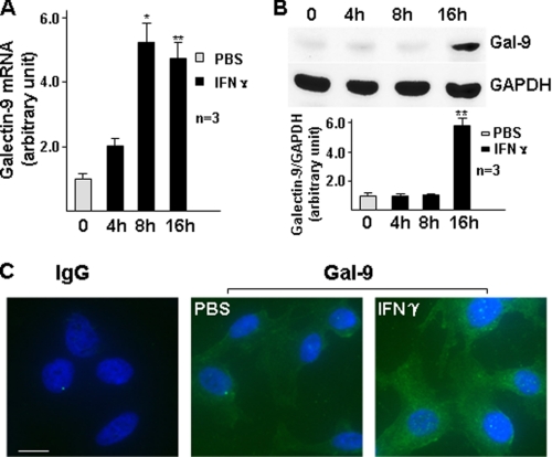
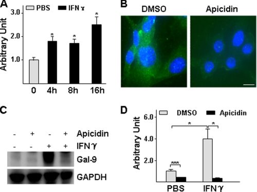
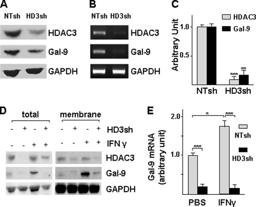
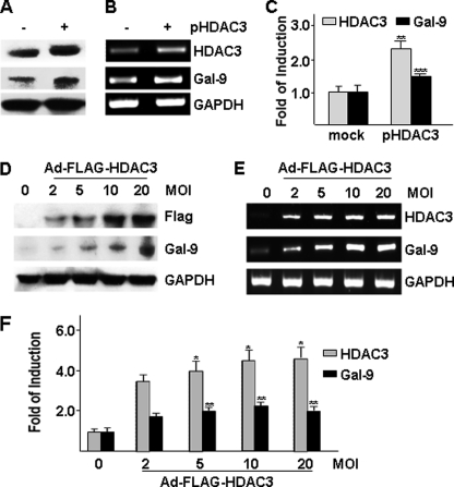
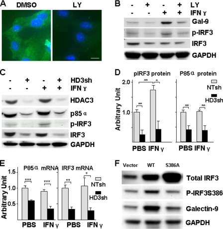
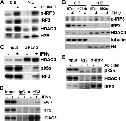
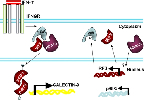
Galectin-9 expression in endothelial cells can be induced in response to inflammation. However, the mechanism of its expression remains unclear. In this study, we found that interferon-γ (IFN-γ) induced galectin-9 expression in human endothelial cells in a time-dependent manner, which coincided with the activation of histone deacetylase (HDAC). When endothelial cells were treated with the HDAC3 inhibitor, apicidin, or shRNA-HDAC3 knockdown, IFN-γ-induced galectin-9 expression was abolished. Overexpression of HDAC3 induced the interaction between phosphoinositol 3-kinase (PI3K) and IFN response factor 3 (IRF3), leading to IRF3 phosphorylation, nuclear translocation, and galectin-9 expression. HDAC3 functioned as a scaffold protein for PI3K/IRF3 interaction. In addition to galectin-9 expression, IFN-γ also induced galectin-9 location onto plasma membrane, which was HDAC3-independent. Importantly, HDAC3 was essential for the constitutive transcription of PI3K and IRF3, which might be responsible for the basal level of galectin-9 expression. The phosphorylation of IRF3 was essential for galectin-9 expression. This study provides new evidence that HDAC3 regulates galectin-9 expression in endothelial cells via interaction with PI3K-IRF3 signal pathway.


Figures

FIGURE 1.
FIGURE 1.
IFN-γ induces galectin-9 expression in HUVECs at mRNA and protein levels. A and B, HUVECs were treated with 10 ng/ml IFN-γ for the time indicated, followed by quantitative RT-PCR analysis of galectin-9 mRNA (A) and Western blot analysis of galectin-9 protein (B, lower panel represents the relative galectin-9 protein level to GAPDH). *, p < 0.05; **, p < 0.01. C, HUVECs were treated with 10 ng/ml IFN-γ for 16 h, followed by immunofluorescent staining with galectin-9 antibody (green). DAPI was included to counterstain the nucleus (blue). PBS was used as vehicle control. IgG was included as primary antibody control of the staining. Scale bar, 5 μm. Note that the fluorescence intensity in IFN-γ-treated cells is significantly higher than untreated cells. Data presented are representative of mean ± S.E. (error bars) of three independent experiments.
FIGURE 2.
FIGURE 2.
HDAC3 activity is essential for galectin-9 expression. A, IFN-γ increases HDAC activity. HUVECs were treated with 10 ng/ml IFN-γ for the time indicated, followed by HDAC activity assay. Arbitrary unit was defined as A420 nm/μg of protein with untreated set as 1.0. *, p < 0.05. B–D, apicidin decreased the basal level and IFN-γ-induced galectin-9 expression. HUVECs were pretreated with 200 nmol/liter apicidin for 1 h, then treated with 10 ng/ml IFN-γ for 16 h in the presence of apicidin, followed by immunofluorescent staining (B: green, galectin-9; blue, DAPI; Scale bar, 5 μm), Western blot assay (C) and quantitative RT-PCR analysis (D) are shown. *, p < 0.05; ***, p < 0.0001. Data presented are representative of mean ± S.E. (error bars) of three independent experiments. DMSO, dimethyl sulfoxide.
FIGURE 3.
FIGURE 3.
Knockdown of HDAC3 decreases basal level and IFN-γ-induced galectin-9 expression. A–C, knockdown of HDAC3 decreased basal level of galectin-9 expression. HUVECs were infected with nontarget (NTsh) or HDAC3 (HD3sh) shRNA lentiviruses at 1 × 10⁷ units/1 × 106 cells for 72 h, followed by Western blotting (A), routine RT-PCR(B), and quantitative RT-PCR (C) assays. An arbitrary unit was defined as the ratio of target gene mRNA to 18 S RNA with that of NTsh set as 1.0. **, p < 0.01; ***, p < 0.0001. D and E, knockdown of HDAC3 abolishes IFN-γ-induced galectin-9 expression. HUVECs were infected with NTsh or HD3sh RNA at 1 × 10⁷ units/1 × 106 cells for 48 h, then treated with 10 ng/ml IFN-γ for 16 h, followed by membrane fraction isolation and RNA extraction. HDAC3 and galectin-9 protein levels were detected in total cell lysate and membrane fractions by Western blotting (D), and mRNA was detected with quantitative RT-PCR (E). *, p < 0.05; ***, p < 0.0001. Data presented are representative of mean ± S.E. (error bars) of three independent experiments.
FIGURE 4.
FIGURE 4.
Overexpression of HDAC3 increases galectin-9 expression. A–C, HUVECs were transfected with pShuttle2-FLAG-HDAC3 plasmid (pHDAC3, 2 μg/2 × 106cells) via electroporation, followed by Western blotting (A), routine RT-PCR (B), and real time RT-PCR (C) assays 48 h after transfection. pShuttle2-FLAG vector was included as mock control. **, p < 0.01; ***, p < 0.0001. D–F, HUVECs were infected with Ad-FLAG-HDAC3 virus at the m.o.i. indicated, followed by Western blotting (D), routine RT-PCR (E), and real time RT-PCR (F) assays 48 h after infection. Ad-null virus was included as negative control and to compensate the m.o.i. *, p < 0.05; **, p < 0.01. -Fold of induction was defined as the ratio of target gene mRNA to 18 S RNA with that of control group set as 1.0. Data presented are representative of mean ± S.E. (error bars) of three independent experiments.
FIGURE 5.
FIGURE 5.
HDAC3 is essential for IFN-γ-induced PI3K-IRF3 activation and galectin-9 expression. A and B, PI3K is crucial for IFN-γ-induced galectin-9 expression and IRF3 activation. HUVECs were pretreated with PI3K inhibitor LY294002 (5 μmol/liter) for 1 h, then treated with 10 ng/ml IFN-γ for 16 h in the presence of LY294002, followed by immunofluorescent staining (A: green, galectin-9; blue, DAPI; Scale bar, 5 μm) and Western blot assay (B). The same amount of dimethyl sulfoxide was included as vehicle control. p-IRF3 indicates phosphorylation of IRF3 at Ser-386. C–E, HDAC3 is crucial for basal level expression of PI3K and IRF3 and IFN-γ-induced IRF3 activation. HUVECs were infected with NTsh or HD3sh RNA for 48 h, then treated with 10 ng/ml IFN-γ for 16 h, followed by Western blot analysis (C, representative image; D, quantitative analysis) and quantitative RT-PCR assays (E). *, p < 0.05; **, p < 0.01. F, IRF3 phosphorylation is essential for galectin-9 expression. HUVECs were transfected with pcDNA3 (vector), pcDNA3-IRF3 (WT), or pcDNA3-IRF3S386A (S386A) plasmids for 48 h, followed by Western blot analysis of IRF3 phosphorylation and galectin-9 expression. Data presented are representative or average ± S.E. (error bars) of three independent experiments.
FIGURE 6.
FIGURE 6.
IFN-γ enhances the HDAC3-p85α-IRF3 complex formation. A, overexpression of HDAC3 induced IRF3 nuclear translocation. HUVECs were infected with 10 m.o.i. Ad-HDAC3 virus for 48 h, followed by cellular fraction assay. Ad-null virus was included as control. C.E, cytosolic extract; N.E, nuclear extract. B, HDAC3 knockdown ablated IFN-γ-induced IRF3 nuclear translocation. HUVECs were infected with NTsh or HD3sh for 72 h, and then treated with 10 ng/ml IFN-γ for 4 h, followed by cellular fraction assay. Anti-tubulin and histone H4 antibodies were included to identify cytosol and nuclear fractions. C, IFN-γ induced the formation of the HDAC3-p85α-IRF3 complex. HUVECs were infected with Ad-FLAG-HDAC3 virus at 10 m.o.i. and 48 h later treated with 10 ng/ml IFN-γ for 4 h, followed by immunoprecipitation assay with anti-FLAG antibody. 50 μg of lysate was included as input. D, IFN-γ enhanced endogenous HDAC3 association with p85α-IRF3. HUVECs were treated with 10 ng/ml IFN-γ for 4 h, followed by immunoprecipitation assay with anti-HDAC3 antibody. E, HDAC3 activity is unnecessary for HDAC3-p85α-IRF3 complex formation. HUVECs were treated with 10 ng/ml IFN-γ for 4 h in the presence or absence of apicidin, followed by immunoprecipitation with anti-IRF3 antibody. Dimethyl sulfoxide was included as vehicle control.
FIGURE 7.
FIGURE 7.
Schematic illustration of IFN-γ-mediated galectin-9 expression in HUVECs. In HUVECs, HDAC3 is involved in the constitutive expression of IRF3 and the p85α subunit of PI3K. Under IFN-γ treatment, HDAC3, PI3K, and IRF3 form a complex, leading to the phosphorylation of IRF3 at serine 386, which in turn activates galectin-9 expression.

References

    1. Leffler H., Carlsson S., Hedlund M., Qian Y., Poirier F. (2004) Glycoconj. J. 19, 433–440 - PubMed
    1. Norling L. V., Perretti M., Cooper D. (2009) J. Endocrinol. 201, 169–184 - PubMed
    1. Yang R. Y., Rabinovich G. A., Liu F. T. (2008) Expert Rev. Mol. Med. 10, e17. - PubMed
    1. Asakura H., Kashio Y., Nakamura K., Seki M., Dai S., Shirato Y., Abedin M. J., Yoshida N., Nishi N., Imaizumi T., Saita N., Toyama Y., Takashima H., Nakamura T., Ohkawa M., Hirashima M. (2002) J. Immunol. 169, 5912–5918 - PubMed
    1. Matsumoto R., Matsumoto H., Seki M., Hata M., Asano Y., Kanegasaki S., Stevens R. L., Hirashima M. (1998) J. Biol. Chem. 273, 16976–16984 - PubMed

Publication types

MeSH terms