Review
. 2012 Jul;32(7):1152-66.
doi: 10.1038/jcbfm.2011.149. Epub 2011 Oct 26.

Sweet sixteen for ANLS

Affiliations
Review

Sweet sixteen for ANLS

Luc Pellerin et al. J Cereb Blood Flow Metab. 2012 Jul.

Abstract

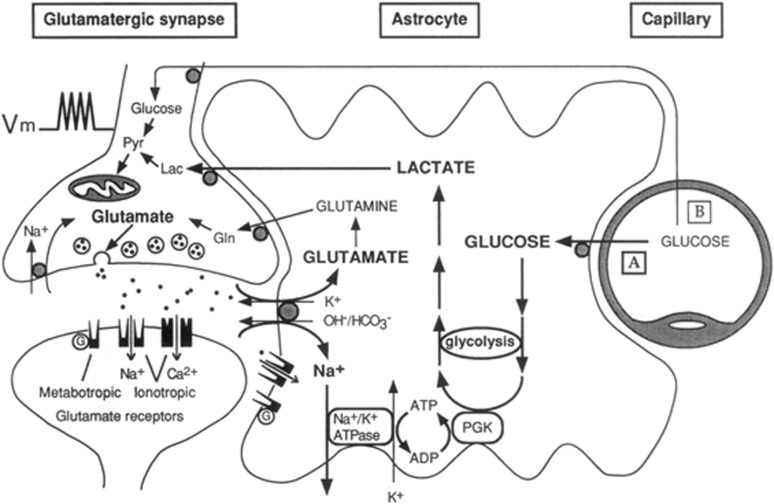
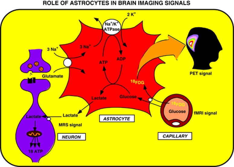
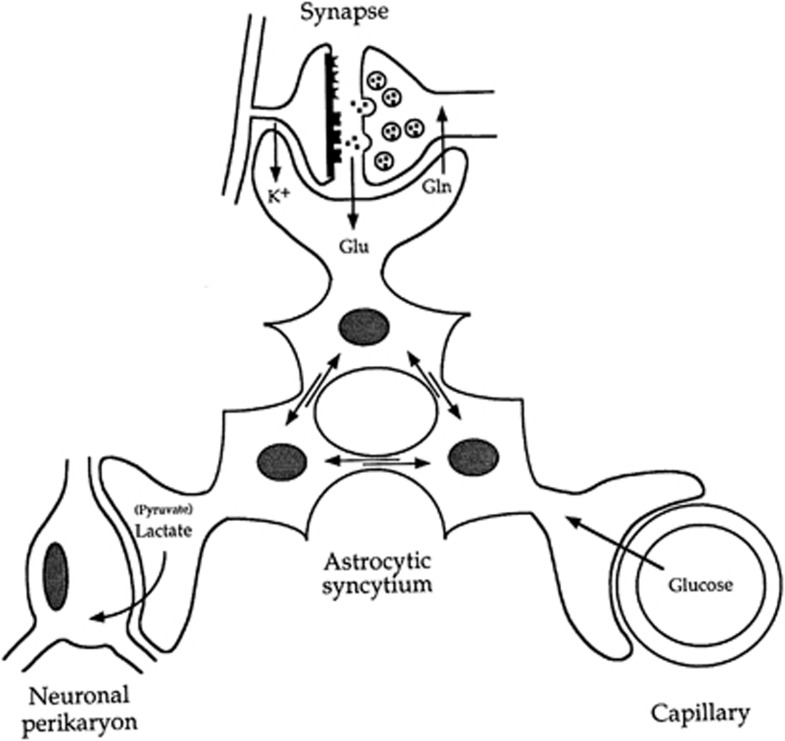
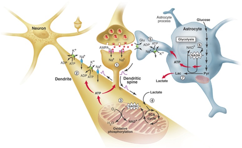
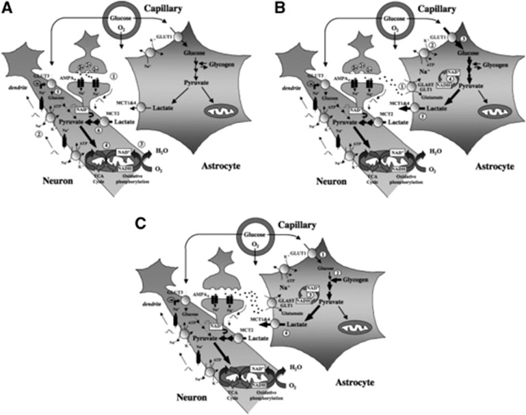
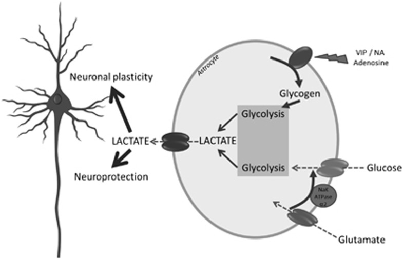
Since its introduction 16 years ago, the astrocyte-neuron lactate shuttle (ANLS) model has profoundly modified our understanding of neuroenergetics by bringing a cellular and molecular resolution. Praised or disputed, the concept has never ceased to attract attention, leading to critical advances and unexpected insights. Here, we summarize recent experimental evidence further supporting the main tenets of the model. Thus, evidence for distinct metabolic phenotypes between neurons (mainly oxidative) and astrocytes (mainly glycolytic) have been provided by genomics and classical metabolic approaches. Moreover, it has become clear that astrocytes act as a syncytium to distribute energy substrates such as lactate to active neurones. Glycogen, the main energy reserve located in astrocytes, is used as a lactate source to sustain glutamatergic neurotransmission and synaptic plasticity. Lactate is also emerging as a neuroprotective agent as well as a key signal to regulate blood flow. Characterization of monocarboxylate transporter regulation indicates a possible involvement in synaptic plasticity and memory. Finally, several modeling studies captured the implications of such findings for many brain functions. The ANLS model now represents a useful, experimentally based framework to better understand the coupling between neuronal activity and energetics as it relates to neuronal plasticity, neurodegeneration, and functional brain imaging.


Figures

Figure 1
Figure 1
The first picture of the astrocyte–neuron lactate shuttle (ANLS) model at birth on 25 October 1994. Schematic of the mechanism for glutamate-induced glycolysis in astrocytes during physiological activation. At glutamatergic synapses, glutamate depolarizes neurons by acting at specific receptor subtypes. The action of glutamate is terminated by an efficient glutamate uptake system located primarily in astrocytes. Glutamate is cotransported with Na+, resulting in an increase in [Na+]i, leading to activation of Na+/K+ ATPase. The pump, fueled by ATP provided by membrane-bound glycolytic enzymes (possibly phosphoglycerate kinase (PGK)), activates glycolysis—that is, glucose utilization and lactate production—in astrocytes. Lactate, once released, can be taken up by neurons and serve as an adequate energy substrate. For graphic clarity, only lactate uptake into presynaptic terminals is indicated. However, this process could also occur at the postsynaptic neuron. Based on recent evidence, glutamate receptors are also shown on astrocytes. This model, which summarizes in vitro experimental evidence indicating glutamate-induced glycolysis, is taken to reflect cellular and molecular events occurring during activation of a given cortical area [arrow labeled A (activation)] direct glucose uptake into neurons under basal conditions is also shown [arrow labeled B (basal conditions)]. Pyr, pyruvate; LAC, lactate; GLN, glutamine; G, guanine nucleotide binding protein. Taken from Pellerin and Magistretti (1994).
Figure 2
Figure 2
Proposed concept derived from the astrocyte–neuron lactate shuttle (ANLS) model that brain imaging signals based on glucose uptake primarily reflect astrocyte metabolism. During synaptic activity, glutamate is released into the synaptic cleft. A glutamate transporter system on astrocytes ensures removal of glutamate from the synaptic cleft. The entry of Na+ cotransported with glutamate activates the Na+/K+ ATPase. Activation of the Na+/K+ ATPase is coupled with an increased glycolytic flux, hence resulting in the stimulation of glucose uptake from the capillaries. Lactate, the major end product of glycolysis, is released by astrocytes and taken up by neurons where it can enter the tricarboxylic acid (TCA) cycle and provide 18 ATP per molecule. This model implies that the activity-linked uptake of 18Fluorodeoxyglucose (18FDG) monitored with positron emission tomography (PET) reflects primarily an astrocyte-based signal. However, since neuronally released glutamate triggers the cascade of events that leads to glucose uptake, the 18FDG-PET signal faithfully reflects activation of neuronal circuits. Taken from Magistretti and Pellerin (1996). fMRI, functional magnetic resonance imaging.
Figure 3
Figure 3
Introduction of the concept of the importance of the astrocytic syncytium for intracellular metabolite trafficking, also known as metabolic polarity of the astrocytic syncytium. Astrocytes have various processes that make contact either with capillaries, neuronal perikarya, or synapses. In addition, they are connected with each other at gap junctions; through these junctions small molecules (MW<1,000) can be exchanged. These properties endow the astrocytic syncytium with the capacity to ensure the transfer of metabolic intermediates from areas of production to areas of demand. Gln, glutamine; Glu, glutamate. Taken from Magistretti et al (1995).
Figure 4
Figure 4
The brain energetics in the limelight. Separate activation of oxidative phosphorylation (respiration) in neurons (brown) and glycolysis in astrocytes (gray), as revealed by two-photon fluorescence imaging of nicotinamide adenine dinucleotide (NADH). (1) Stimulation of excitatory (glutamatergic) neurons activates postsynaptic AMPA receptors and induces an excitatory postsynaptic potential (EPSP) in the dendritic spine of the neuron. (2) The depolarization propagates from the dendritic spine to the dendrite, where it may cause further opening of voltage-gated sodium channels and activation of the Na+/K+ ATPase, leading to an increased demand for energy (ATP). (3) In response, oxidative phosphorylation is rapidly activated, causing a decrease in mitochondrial NADH content (the so-called ‘dip' in the fluorescent signal). (4) Recovery of mitochondrial NADH in dendrites is accomplished by stimulation of the tricarboxylic acid (TCA) cycle, fueled largely by lactate from the extracellular pool. (5) In parallel, but delayed in time, glutamate reuptake in astrocytes (gray) activates the glial Na+/K+ ATPase. (6) The increased energy demand leads to a strong enhancement of glycolysis in the cytoplasm of astrocytes, as indicated by the large increase in cytosolic NADH fluorescence (the so-called ‘overshoot'). (7) To maintain the high glycolytic flux, NAD+ must be regenerated via the conversion of pyruvate to lactate through the activity of the enzyme lactate dehydrogenase. Release of lactate into the extracellular space not only replenishes the extracellular pool, but also may sustain the late phase of neuronal activation. In vivo, glucose is delivered from the blood to both the extracellular space and astrocytes (via astrocytic protrusions called end-feet that are in close contact with the blood vessel wall). AMPAR, α-amino-3-hydroxy-5-methyl-4-isoxazole propionic acid receptors; Glu, glutamate; Lac, lactate; Pyr, pyruvate. Taken from Pellerin and Magistretti (2005).
Figure 5
Figure 5
Importance of monocarboxylate transporters and the regulation of their expression/localization in lactate shuttling between astrocytes and neurons. Several unique features of astrocytes and neurons have been uncovered that suggest a partial metabolic compartmentalization and the existence of a preferential lactate transfer between the two cell types. Thus, exposure of astrocytes to glutamate was shown to directly enhance glucose transport in parallel with increased glucose utilization. Conversion of pyruvate into lactate in astrocytes is facilitated by key characteristics. Astrocytes lack a mitochondrial aspartate/glutamate carrier that reduces their capacity to transfer reducing equivalent as nicotinamide adenine dinucleotide (NADH) by the malate/aspartate shuttle in the mitochondria and regenerate NAD. To maintain the glycolytic flux, cytosolic NADH is rather converted to NAD through the reaction catalyzed by the lactate dehydrogenase isoform LDH5, preferentially expressed in astrocytes. Lactate formed is then released in the extracellular space via the high-capacity monocarboxylate transporters, MCT1 and MCT4. In contrast to astrocytes, glucose uptake in neurons is reduced by glutamate. Moreover, ascorbic acid that is shuttled from astrocytes to neurons also contributes to reduce glucose uptake in neurons. In parallel, ascorbic acid enhances lactate uptake by neurons, and lactate conversion to pyruvate is facilitated by the preferential expression of the lactate dehydrogenase isoform, LDH1. Expression of the neuronal monocarboxylate transporter, MCT2, can be enhanced through an increase in protein synthesis by various stimuli, which might contribute to long-term adaptation of energy supply to demand. GLUT, glucose transporter; Lac, lactate; LDH, lactate dehydrogenase; MCT, monocarboxylate transporter; Pyr, pyruvate. Taken from Pellerin (2008).
Figure 6
Figure 6
Contribution of glucose- and glycogen-derived lactate to support different phases of neuronal activation. (A) Early phase. Activation of glutamatergic afferents leads to synaptic release of glutamate, AMPA receptor (AMPAR) activation, and generation of an excitatory postsynaptic potential (EPSP) caused by Na+ entry within the postsynaptic spine (1). Depolarization propagates to the dendrite and causes opening of voltage-sensitive Na+ channels, leading to further Na+ entry. Reestablishment of ion gradients is accomplished by the Na+/K+ ATPase (2), which creates considerable energy expenditure. As a consequence, oxidative phosphorylation is activated (3) and mitochondrial nicotinamide adenine dinucleotide (NADH) levels first decrease (Kasischke et al, 2004). Then, enhanced tricarboxylic acid (TCA) cycle activity will ensue (4) to supply NADH for oxidative phosphorylation and support ATP production. As pyruvate utilization in the TCA cycle increases and its cytoplasmic levels decrease, the conditions become favorable for both enhanced glucose and lactate use. Surprisingly, activation of AMPA receptors and coupled Na+ entry lead to a reduction in glucose uptake and utilization in neurons (5), thus further favoring lactate utilization as preferential oxidative substrate (6). This would cause a transient drop in extracellular lactate levels as measured in vivo. (B) Late phase. Glutamate released in the synaptic cleft is taken up by astrocytes to be recycled via the specific glutamate transporters GLAST and GLT1 (1). A large Na+ influx caused by glutamate uptake takes place and activates the Na+/K+ ATPase (2), glucose transport (3) and (4) glucose utilization in astrocytes. The enhancement of aerobic glycolysis in astrocytes first causes a large increase in cytosolic NADH that normalizes with the conversion of pyruvate into lactate and its release via monocarboxylate transporters expressed on astrocytes (mainly MCT1 and 4) (5). Such a lactate release following glutamatergic activation corresponds to the increase in extracellular lactate levels measured in vivo. Lactate produced by astrocytes during this later phase of activation not only replenishes the extracellular pool but also could help sustain neuronal energy needs as activation persists. Metabolic events occurring in the early and late phases described above constitute the so-called astrocyte–neuron lactate shuttle and its importance grows with the degree of glutamatergic activation. Such a view is supported by a series of experiments conducted in vivo. (C) Intense and prolonged stimulation. On strong and long-lasting stimulation that occurs in certain conditions, glucose utilization becomes very important, in part due to intense glutamate reuptake in astrocytes, that extracellular glucose levels are insufficient to sustain such uptake (1). In such a situation, glycogen present in the astrocyte is mobilized to provide the necessary glycosyl units (2) as previously demonstrated in vivo. Glycolysis is the predominant pathway (3) and lactate is produced (4) to maintain the high glycolytic rate. Resynthesis of glycogen will cause additional glucose uptake that might contribute to create a mismatch between glucose utilization and oxygen consumption, a phenomenon known as ‘uncoupling.' Taken from Pellerin et al (2007).
Figure 7
Figure 7
Complementary spatial domains in which vasoactive intestinal peptide (VIP)-containing bipolar neurons and noradrenergic fibers exert their glycolytic effects. Such an arrangement would allow for the establishment of temporary columnar ‘hotspots.' VIP, VIP-containing bipolar cell; NA, noradrenergic afferents; Pyr, pyramidal cell furnishing major efferent projections; SA, specific afferent (i.e., from thalamus or from other cortical areas); WM, subcortical white matter. Cortical layers denoted by roman numerals. Note tangential orientation of NA fibers, and radially restricted domain of VIP-containing neuron. The concomitant activation of noradrenergic fibers (large, yellow arrow, bottom) by unexpected sensory stimuli and of a group of VIP-containing intracortical neurons by specific thalamic inputs (small, blue arrow, bottom left) would lead to a drastic increase in cAMP levels within a discrete volume of somatosensory cortex, delineated here by the cylinder with dark background. Adjoining gray-background cylinders represent nonactivated cortical volumes. For graphic clarity, only the VIP-containing (green) and the pyramidal cells (red) have been represented here. In particular, the principal target cells of the thalamocortical afferents, that is, the small stellate cells in layer IV, have been omitted. However, any cell with the capacity for dendritic reception in layer IV may receive thalamic inputs. Furthermore, also for graphic clarity, only one VIP neuron per cylinder has been drawn, in representation of a group of VIP neurons. This is a heuristic model, and as such, certain details of the synaptic circuitry depicted in the diagram (e.g., direct thalamocortical input to VIP-containing cells) are suggested by presently available data, but have not been demonstrated definitively. However, all aspects of the model are potentially testable. Taken from Magistretti and Morrison (1988).
Figure 8
Figure 8
Roles of glucose- and glycogen-derived lactate in neuroprotection and neuronal plasticity. It is suggested that under different levels of brain activation, glutamatergic as well as specific neuromodulatory systems (e.g., noradrenergic) will be activated. Glutamate reuptake in astrocytes will trigger glucose-derived lactate production and release. In parallel, neuromodulators such as vasoactive intestinal peptide (VIP), noradrenaline (NA), or adenosine will stimulate glycogenolysis, leading as well to lactate production and release. Such lactate provision to neurons by astrocytes turns out to be essential for the establishment of memory via support of synaptic plasticity processes as well as for neuroprotection of neurons under certain stress conditions (e.g., excitotoxicity).

References

    1. Ainscow EK, Mirshamsi S, Tang T, Ashford ML, Rutter GA. Dynamic imaging of free cytosolic ATP concentration during fuel sensing by rat hypothalamic neurones: evidence for ATP-independent control of ATP-sensitive K(+) channels. J Physiol. 2002;544:429–445. - PMC - PubMed
    1. Allen NJ, Káradóttir R, Attwell D. A preferential role for glycolysis in preventing the anoxic depolarization of rat hippocampal area CA1 pyramidal cells. J Neurosci. 2005;25:848–859. - PMC - PubMed
    1. Andriezen WL. On a system of fibre-like cells surrounding the blood vessels of the brain of man and mammals, and its physiological significance. Int Monatsschr Anat Physiol. 1893;10:532–540.
    1. Attwell D, Laughlin SB. An energy budget for signaling in the grey matter of the brain. J Cereb Blood Flow Metab. 2001;21:1133–1145. - PubMed
    1. Aubert A, Costalat R, Magistretti PJ, Pellerin L. Brain lactate kinetics: modeling evidence for neuronal lactate uptake upon activation. Proc Natl Acad Sci USA. 2005;102:16448–16453. - PMC - PubMed

Publication types

LinkOut - more resources