. 2012 Apr;22(4):721-30.
doi: 10.1007/s00330-011-2312-2. Epub 2011 Oct 26.

Real-time tissue elastography for testicular lesion assessment

Affiliations

Real-time tissue elastography for testicular lesion assessment

Alfredo Goddi et al. Eur Radiol. 2012 Apr.

Abstract

Objectives: To assess the ability of Real-time Elastography (RTE) to differentiate malignant from benign testicular lesions.

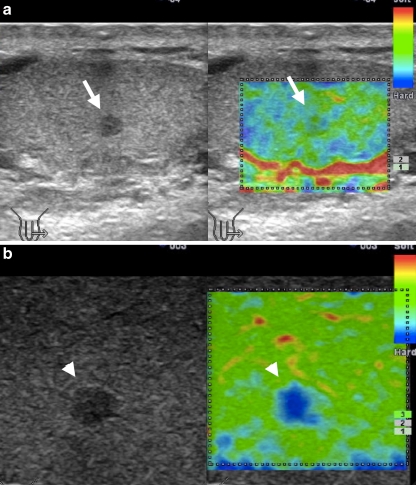
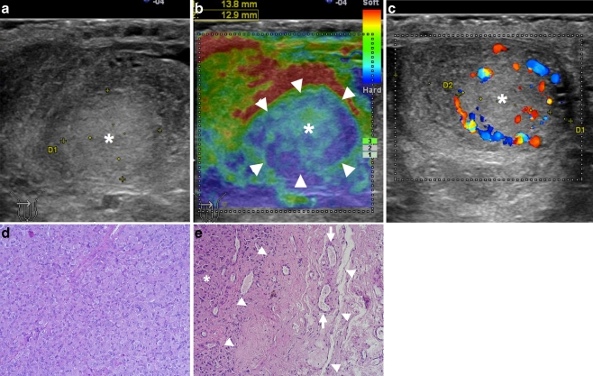
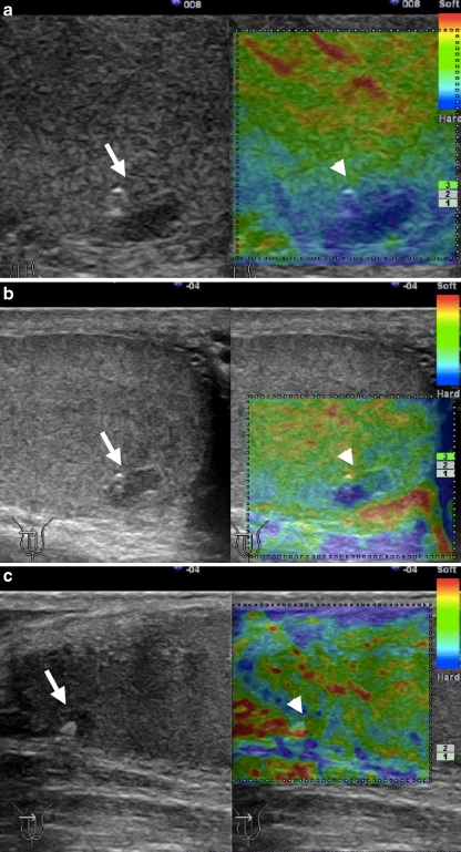
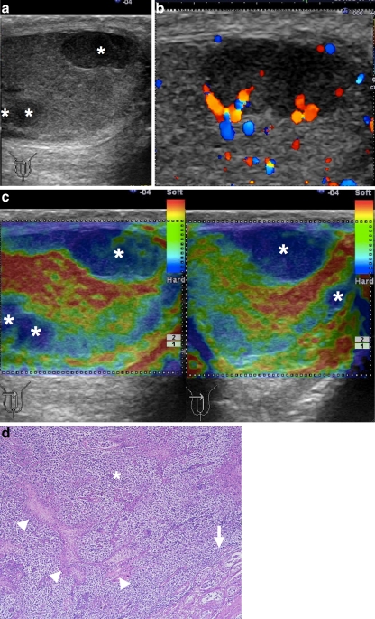
Methods: In 88 testicles ultrasound identified 144 lesions, which were examined by RTE. Elasticity images of the lesions were assigned the colour-coded score of Itoh (Radiology 2006), according to the distribution of strain induced by light compression. RTE findings were analysed considering shape (nodular/pseudo-nodular), size (<5 mm, 6-10 mm, >11 mm) and score (SC1-5) of the lesions.

Results: 93.7% of all benign lesions showed a complete elastic pattern (SC1). 92.9% of benign nodules <5 mm and 100% of the pseudonodules showed a nearly complete elastic pattern (mainly SC1). 87.5% of malignant nodules showed a stiff pattern (SC4-5). RTE gave 87.5% sensitivity, 98.2% specificity, 93.3% positive predictive value, 96.4% negative predictive value and 95.8% accuracy in differentiating malignant from benign lesions.

Conclusions: RTE is a useful technique in assessing small testicular nodules and pseudo-nodules. This is relevant in clinical practice allowing expectant management in RTE selected cases. The role of RTE seems less relevant for larger lesions because most of them are malignant at clinical and ultrasound assessment, limiting RTE to simply confirmation role.

Key points: An emerging role for Elastography in allowing surveillance for small testicular lesions. Elastography can better differentiate benign from malignant testicular lesions. Follow up can be reduced for elastic testicular lesions at Elastography.


Figures

Fig. 1
Fig. 1
a Grey-scale ultrasound longitudinal image of the testis: normal homogeneous fine granular echo pattern. b Elastography: a wide RTE box was selected to evaluate testicular elasticity. The typical distribution of elasticity in a normal testis shows mainly green (intermediate), some linear red (soft) (arrowheads) and peripheral light blue (stiff); outside the testicle RTE shows red (soft) related to the fluid in the intravaginal space (arrow). On the right side the quality scale indicating levels 3
Fig. 2
Fig. 2
a Case # 1: Ultrasound showed a small hypoechoic nodule (3 mm in size), with homogeneous elasticity (score 1) at RTE (arrow). Follow-up highlighted the ability of RTE to assess the benign aspect. b Case # 2: a 4-mm hypoechoic nodule at ultrasound, completely anaelastic (score 4) at RTE (arrowhead), was considered suspicious. A close follow-up carried out at regular intervals for 30 months showed unchanged size and appearance. The lesion has therefore been reclassified as “probably benign” and its inelastic aspect as a false-positive of RTE
Fig. 3
Fig. 3
a Ultrasound showed a nodule (14 mm in size) slightly hyperechoic (*) compared with the testicular tissue. b Unusual appearance on RTE: the central strain area (*) was surrounded by a nearly complete no strain – stiff – rim (arrowheads; i.e. the peripheral part of the lesion was blue, the central part was green). It was must classified as Score 2. The relative elasticity (i.e. displacement) of different tissue showed by RTE, explains the apparent softer aspect of the anterior portion of the rim (displayed in light blue instead of dark blue) and of the adjacent testicle (displayed in red). This is the consequence of the higher tissue displacement generated in the near field by the applied compressional forces. c Colour Doppler showed mainly peripheral vascularisation. d Pathological specimens of the testes: Leydig tumour. Central area of the nodule: polygonal cells with abundant, eosinophilic cytoplasm, solid, sheet-like pattern and no interstitial fibrous stroma. Haematoxylin-Eosin (×100). e Peripheral area of the nodule: sclerohyalinosis of the testicular tissue (arrowheads) adjacent to the neoplasm (*); peripheral vessels surrounding the tumor are visibile (arrows; ×100)
Fig. 4
Fig. 4
a Ultrasound showed a subcapsular oval-shaped hypoechoic pseudonodule (arrow) and an adjacent rounded hyperechoic spot in a patient with recent scrotal trauma. At RTE the entire lesion and its surrounding area were blue (arrowhead), indicating a score 5 appearance that usually indicates suspected neoplasm. Considering the shape and the recent history of trauma, a close follow-up was planned. b Six months later the B-mode image seemed to be about the same (arrow), but at RTE half of the nodule showed regular strain and the surrounding stiffness had disappeared (arrowhead). c After 9 months it was reduced in size, partially calcified (arrow) and became elastic (score 1; arrowhead)
Fig. 5
Fig. 5
a Multiple hypoechoic testicular nodules at ultrasound (*). b Colour Doppler of the largest nodule showed mainly peripheral vascular signals. c Longitudinal and transverse testicular imaging. The nodules (*), score 4–5 at RTE, were correctly identified as tumours. d Pathological specimen. Multifocal seminoma of the testis at histological diagnosis: high cellularity, typical seminoma cells (*) with discrete interstitial stroma (arrowheads) and thick fibrous pseudocapsule (arrow; ×40)

Comment in

References

    1. Ragheb D, Higgins JL., Jr Ultrasonography of the scrotum: technique, anatomy and pathologic entities. J Ultrasound Med. 2002;21:171–185. - PubMed
    1. Mihmanli I, Kantarci F. Sonography of scrotal abnormalities in adults: an update. Diagn Interv Radiol. 2009;15:64–73. - PubMed
    1. Krouskop TA, Dougherty DR, Vinson FS. A pulsed Doppler ultrasonic system for making noninvasive measurements of the mechanical properties of soft tissue. J Rehabil Res Dev. 1987;24:1–8. - PubMed
    1. Ophir J, Céspedes I, Ponnekanti H, Yazdi Y, Li X. Elastography: a quantitative method for imaging the elasticity of biological tissues. Ultrason Imaging. 1991;13:111–134. doi: 10.1016/0161-7346(91)90079-W. - DOI - PubMed
    1. Shiina T, Nitta N, Ueno E, Bamber JC. Real time tissue elasticity imaging using the combined autocorrelation method. J Med Ultrasonics. 2002;29:119–128. doi: 10.1007/BF02481234. - DOI - PubMed