. 2012 Feb;27(2):374-89.
doi: 10.1002/jbmr.548.

Cell autonomous requirement of connexin 43 for osteocyte survival: consequences for endocortical resorption and periosteal bone formation

Affiliations

Cell autonomous requirement of connexin 43 for osteocyte survival: consequences for endocortical resorption and periosteal bone formation

Nicoletta Bivi et al. J Bone Miner Res. 2012 Feb.

Abstract

Connexin 43 (Cx43) mediates osteocyte communication with other cells and with the extracellular milieu and regulates osteoblastic cell signaling and gene expression. We now report that mice lacking Cx43 in osteoblasts/osteocytes or only in osteocytes (Cx43(ΔOt) mice) exhibit increased osteocyte apoptosis, endocortical resorption, and periosteal bone formation, resulting in higher marrow cavity and total tissue areas measured at the femoral mid-diaphysis. Blockade of resorption reversed the increased marrow cavity but not total tissue area, demonstrating that endocortical resorption and periosteal apposition are independently regulated. Anatomical mapping of apoptotic osteocytes, osteocytic protein expression, and resorption and formation suggests that Cx43 controls osteoclast and osteoblast activity by regulating osteoprotegerin and sclerostin levels, respectively, in osteocytes located in specific areas of the cortex. Whereas empty lacunae and living osteocytes lacking osteoprotegerin were distributed throughout cortical bone in Cx43(ΔOt) mice, apoptotic osteocytes were preferentially located in areas containing osteoclasts, suggesting that osteoclast recruitment requires active signaling from dying osteocytes. Furthermore, Cx43 deletion in cultured osteocytic cells resulted in increased apoptosis and decreased osteoprotegerin expression. Thus, Cx43 is essential in a cell-autonomous fashion in vivo and in vitro for osteocyte survival and for controlling the expression of osteocytic genes that affect osteoclast and osteoblast function.


Figures

Figure 1
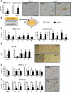
Figure 1. Deletion of Cx43 from osteocytes and osteoblasts results in increased cortical osteocyte apoptosis, endocortical osteoclastic surface and periosteal apposition
(A) Apoptotic (TUNEL positive) osteocytes were scored in the cortical bone of femora, 2.5 mm from the midshaft towards the growth plate. Representative images show examples of living (black arrows) and apoptotic (red arrows) osteocytes. ALN: alendronate. nd: non-detectable. (B) Representative TEM images of osteocytes in femoral midshaft. (C) Osteoclast number and surface were determined on the endocortical surface of the femoral midshaft. Representative images of bone sections stained for TRAP. Arrows point to TRAP positive osteoclasts. (D and E) Dynamic histomorphometric parameters were measured on the periosteal (D) and endocortical (E) surfaces of the femoral midshaft. ALN: alendronate. (F) Circulating levels of osteocalcin (OCN) measured in plasma. Bars are mean ± SD. * indicates significant differences versus vehicle-treated Cx43fl/- mice and # indicates significant differences versus vehicle-treated Cx43ΔOb-Ot/- mice at p<0.05, n = 6-9.
Figure 2
Figure 2. Effective removal of Cx43 from osteocytes but not from osteoblasts in DMP1-8Kb-Cre;Cx43fl/fl (Cx43ΔOt) mice
(A) Cre mRNA expression in brain, kidney, heart, skeletal muscle and tibia isolated from Cx43fl/fl and Cx43ΔOt mice. (B) Genomic DNA was purified from the indicated tissues and PCR reactions for the deleted allele of Cx43 (Cx43del) and for endogenous DMP1 used as loading control. (C) mRNA expression of keratocan, SOST, and Cx43 was determined in cortical bone preparations before (left panels), and after (right panels) enzymatic digestion with collagenase in order to remove remaining osteoblasts to obtain osteocyte-enriched bone preparations. Bars are mean ± SD of five determinations (3-5 mice/preparation). * indicates significant differences versus Cx43fl/fl mice at p<0.05. (D) Messenger RNA expression in primary osteocytes and osteoblasts isolated from DMP1-8kb-GFP mice expressing Cre recombinase under the control of the 8kb fragment of the DMP1 promoter. OT: osteocytic cells; OB: osteoblastic cells. nd: non-detectable. Bars are mean ± SD of triplicate determinations. (E) Cx43 (brown) expression in femoral cortical bone of Cx43fl/fl and Cx43ΔOt mice stained with an anti-Cx43 polyclonal antibody or non-immune (n.i.) IgG, and counterstained with 0.2% methyl green to reveal cell nuclei (green). Red arrows point to osteocytes and black arrows point to osteoblasts. Representative sections from 3 different animals per genotype are shown. Bar indicates 20 μm.
Figure 3
Figure 3. Removal of Cx43 in Cx43ΔOt mice causes increased osteocyte apoptosis localized to the posterior portion of the femoral cortex, accompanied by targeted osteoclast recruitment
(A) Apoptotic cells were visualized by TUNEL staining (brown) in the total cortical area in sections counterstained with methyl green (green) to detect viable cells. Percentages were calculated over the sum of viable osteocytes, apoptotic osteocytes and empty lacunae. Representative images of the posterior side in specimens stained for TUNEL are shown. Higher magnification image of TUNEL positive apoptotic osteocytes showing abundant TUNEL signal within the canalicular system (red arrow), indicating the presence of fragmented DNA. Bars indicate 20 μm. (B) Schematic indicates the areas in which the cortical bone was divided to measure the parameters showed in this figure. Measurements were made in two equally separated halves of the cortex: periosteal region (Ps) and endocortical region (Ec). Cells were also evaluated around the longitudinal axis in four different portions: anterior (A), lateral (L), posterior (P), and medial (M). (C) Prevalence of osteocyte apoptosis and empty lacunae in the two halves and in the four anatomical regions of the cortex. Bars are mean ± SD. * indicates significant differences versus Cx43fl/fl mice at p<0.05, n=3-5. (D) Osteoclast number on the endocortex (Ec) was determined in sections stained for TRAP and counterstained with Toluidine blue. Representative images of the posterior side stained for TRAP (arrow) are shown (400X). (E and F) The prevalence of RANKL-expressing (E) and OPG-expressing (F) osteocytes was quantified in sections stained with specific antibodies. Bars are mean ± SD. * indicates significant differences at p<0.05 versus Cx43fl/fl mice, n=3-5. Representative images of femoral cross-sections stained with antibodies directed against RANKL (E) and OPG (F) in Cx43fl/fl mice. Bar indicates 20 μm.
Figure 4
Figure 4. Expression of Cx43 in osteocytes is required in a cell autonomous manner to maintain osteocyte viability and a low RANKL/OPG ratio in vitro
(A) Cell number, viability, and proliferation were evaluated by MTT, Trypan blue uptake, and BrdU, respectively 1, 2 and 3 days after seeding in MLO-Y4 cells silenced with scrambled or Cx43 shRNA. Each point corresponds to the mean ± SD of 6-12 replicas. * indicates significant differences versus scramble shRNA-infected cells for each time point at p<0.05. (B) Cx43 protein levels were quantified by Western blotting, corrected by ERK1/2 levels. A representative Western blot image is shown. mRNA levels for Cx43, RANKL and OPG relative to the housekeeping gene Mrps2 were determined by qPCR in MLO-Y4 expressing or not Cx43. Bars are mean ± SD of triplicate determinations. * indicates significant differences at p<0.05 versus scramble shRNA-infected cells. (C) mRNA levels for Cx43, RANKL and OPG relative to the housekeeping gene GAPDH were determined by qPCR in calvaria cells isolated from control Cx43fl/fl and Cx43ΔOt mice. Bars are mean ± SD of quadruplicate determinations. * indicates significant differences at p<0.05 versus control. (D) mRNA levels for Cx43 and SOST relative to the housekeeping gene β-actin were determined by qPCR in Cx43-transfected (UMR+43) and wild type UMR-106 cells. Bars are mean ± SD of triplicate determinations. * indicates significant differences at p<0.05 versus wild type cells.
Figure 5
Figure 5. Increased bone apposition in Cx43ΔOt mice coincides with surfaces adjacent to areas where sclerostin expression is low
(A) Dynamic histomorphometric parameters were measured on the periosteal surface of the femoral midshaft. Bars are mean ± SD. * indicates significant differences at p<0.05 versus Cx43fl/fl mice, n=7-8. Representative images of calcein labels are shown. Left, entire cross-section at low magnification (50X), bar indicates 500 μm; right, high magnification showing the detail of the double labeling on the posterior region. Arrows point to the periosteal surface. Dynamic histomorphometric parameters in the anterior (A), lateral (L), posterior (P), and medial (M) regions around the femoral midshaft, on the periosteal surface. The prevalence of sclerostin (+) osteocytes was determined in the periosteal (Ps) half as a whole (total) and divided in the four anatomical regions, stained with anti-sclerostin antibodies and counterstained with methyl green. Bars are mean ± SD. * indicates significant differences at p<0.05 versus Cx43fl/fl mice, n=3-8. (B) Dymamic histomorphometric parameters were measured on the endocortical surface as a whole and divided in the four anatomical regions. Sclerostin (+) osteocytes were quantified in the endocortical half (Ec) of the bone as a whole (total) and divided in the four anatomical regions. Arrows point to the endocortical surface. Bars are mean ± SD. * indicates significant differences at p<0.05 versus Cx43fl/fl mice, n=3-8.
Figure 6
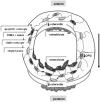
Figure 6. Cx43 deficiency increases osteocyte apoptosis and modulates the levels of osteocytic genes that affect osteoclast and osteoblast function resulting in altered bone geometry
Deletion of Cx43 increases apoptosis and reduces OPG gene expression in osteocytes, inducing osteoclast recruitment and resorption in the posterior endocortical area. Accumulation of empty lacunae causes a reduction in sclerostin levels with consequent increase in bone formation in the anterior endocortical surface and in the posterior periosteal surface. This results in magnification of the physiological modeling drift (represented by the arrow) by which long bones adapt their shape to mechanical needs.

References

    1. Klein-Nulend J, Bonewald LF. The osteocyte. In: Bilezikian JP, Raisz LG, Martin TJ, editors. Principles of Bone Biology. Third ed. Academic Press; San Diego, CA: 2008. pp. 153–174.
    1. Paszty C, Turner CH, Robinson MK. Sclerostin: a gem from the genome leads to bone-building antibodies. J Bone Miner Res. 2010;25:1897–1904. - PubMed
    1. Zhao S, Zhang YK, Harris S, Ahuja SS, Bonewald LF. MLO-Y4 osteocyte-like cells support osteoclast formation and activation. J Bone Miner Res. 2002;17:2068–2079. - PubMed
    1. Kramer I, Halleux C, Keller H, Pegurri M, Gooi JH, Weber PB, Feng JQ, Bonewald LF, Kneissel M. Osteocyte Wnt/beta-catenin signaling is required for normal bone homeostasis. Mol Cell Biol. 2010;30:3071–3085. - PMC - PubMed
    1. O'Brien CA, Plotkin LI, Galli C, Goellner J, Gortazar AR, Allen MR, Robling AG, Bouxsein M, Schipani E, Turner CH, Jilka RL, Weinstein RS, Manolagas SC, Bellido T. Control of bone mass and remodeling by PTH receptor signaling in osteocytes. PLoS ONE. 2008;3:e2942. - PMC - PubMed

Publication types

MeSH terms