. 2012 Jan;5(1):73-82.
doi: 10.1242/dmm.008128. Epub 2011 Oct 25.

Tracing notochord-derived cells using a Noto-cre mouse: implications for intervertebral disc development

Affiliations

Tracing notochord-derived cells using a Noto-cre mouse: implications for intervertebral disc development

Matthew R McCann et al. Dis Model Mech. 2012 Jan.

Abstract

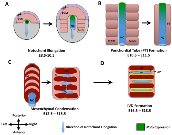
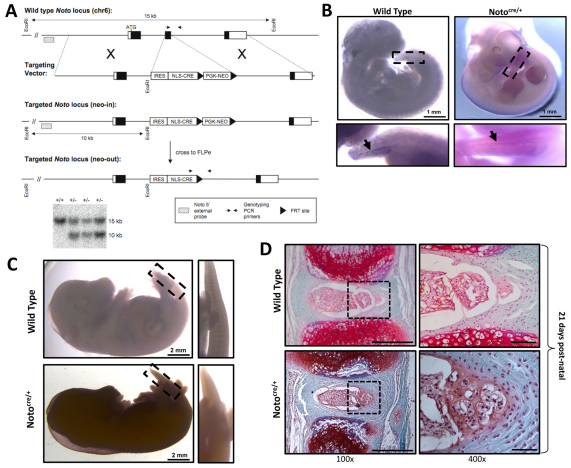
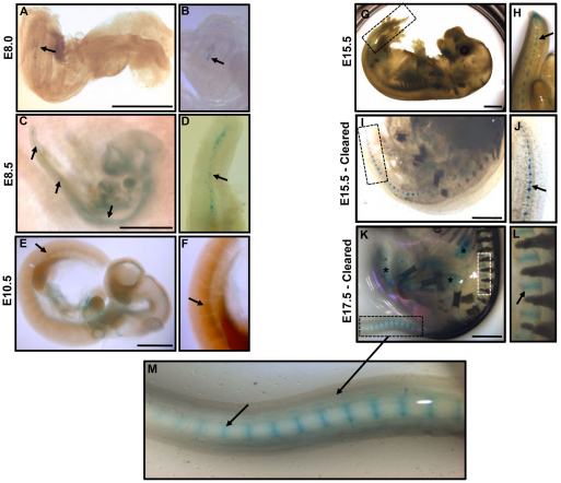
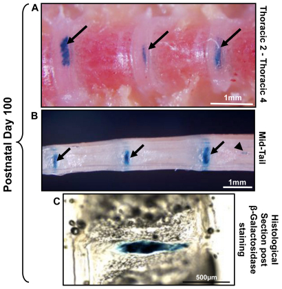
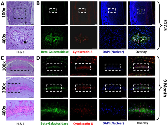
Back pain related to intervertebral disc degeneration is the most common musculoskeletal problem, with a lifetime prevalence of 82%. The lack of effective treatment for this widespread problem is directly related to our limited understanding of disc development, maintenance and degeneration. The aim of this study was to determine the developmental origins of nucleus pulposus cells within the intervertebral disc using a novel notochord-specific Cre mouse. To trace the fate of notochordal cells within the intervertebral disc, we derived a notochord-specific Cre mouse line by targeting the homeobox gene Noto. Expression of this gene is restricted to the node and the posterior notochord during gastrulation [embryonic day 7.5 (E7.5)-E12.5]. The Noto-cre mice were crossed with a conditional lacZ reporter for visualization of notochord fate in whole-mount embryos. We performed lineage-tracing experiments to examine the contribution of the notochord to spinal development from E12.5 through to skeletally mature mice (9 months). Fate mapping studies demonstrated that, following elongation and formation of the primitive axial skeleton, the notochord gives rise to the nucleus pulposus in fully formed intervertebral discs. Cellular localization of β-galactosidase (encoded by lacZ) and cytokeratin-8 demonstrated that both notochordal cells and chondrocyte-like nucleus pulposus cells are derived from the embryonic notochord. These studies establish conclusively that notochordal cells act as embryonic precursors to all cells found within the nucleus pulposus of the mature intervertebral disc. This suggests that notochordal cells might serve as tissue-specific progenitor cells within the disc and establishes the Noto-cre mouse as a unique tool to interrogate the contribution of notochordal cells to both intervertebral disc development and disc degeneration.


Figures

Fig. 1.
Fig. 1.
Schematic representation of the main stages in axial skeletogenesis. (A) Formation of the node (N) and elongation of the notochord (NC). LPM, lateral plate mesoderm; PXM, paraxial mesoderm. (B) Aggregation of the somatic mesenchyme around the notochord leads to formation of a continuous perichordal tube (PT). Localization of Noto expression at these time points is indicated in green. (C) Condensation of the axial mesenchyme leads to spine segmentation and perichordal disc formation. AF, annulus fibrosis; VB, vertebral bodies. (D) Formation of intervertebral discs (IVDs) is associated with the disappearance of the notochord within the vertebral bodies, and its expansion within the IVD. CEP, cartilage end-plates; NP, nucleus pulposus.
Fig. 2.
Fig. 2.
Generation of the notochord-specific Cre mouse line. (A) Targeting strategy used to generate the Notocre/+ line. An internal ribosome entry site-nuclear localization signal-Cre recombinase (IRES-NLS-CRE) cassette replaced exon 2 of the Noto locus. Positively targeted ES cell clones were confirmed by Southern blot using an external 5′ probe; the wild-type allele is 15 kb and the targeted allele is 10 kb. Representative positive ‘neo-in’ clones are shown. The positions of genotyping PCR primers for wild-type and ‘neo-out’ mice are also shown. (B) Targeting of the Noto locus does not affect its temporal regulation, as demonstrated by whole mount in situ hybridization at E11.5. Noto expression is detected in both Notocre/+ and wild-type (Noto+/+) littermate control embryos, localized to the posterior node in the tail region (insert and arrows). (C) Noto expression is downregulated after E12.5, confirmed by in situ hybridization at E15.5 showing no detectable Noto expression. (D) Heterozygous inactivation of Noto does not disrupt notochord formation or IVD development. IVD formation and tissue architecture was examined in Notocre/+ mice and wild-type (Noto+/+) littermate controls using Safranin-O/Fast Green staining on paraffin embedded sections at P21. Enlarged view of the NP and inner AF tissues are shown in the right-hand box. Scale bars: 500 μm for 100× images and 50 μm for 400× images.
Fig. 3.
Fig. 3.
Whole-mount examination of β-galactosidase staining in Notocre/+;R26R/+ embryos. (A–M) Staining pattern of β-galactosidase, demonstrating the localization of notochord-derived cells throughout embryonic development (stages indicated). Higher-magnification view of β-galactosidase expression at each respective time point is presented in panels to the right. Arrows denote positive β-galactosidase staining at each embryonic stage. (I–M) β-galactosidase expression in E15.5 and E17.5 embryos, rendered clear to enable tissue visualization, shows expression within IVDs in the developing spine. Asterisks (*) indicate non-specific background staining; specificity of Cre expression was confirmed by comparing background staining in Noto+/+ littermate controls (data not shown). Scale bars: 1 mm.
Fig. 4.
Fig. 4.
β-galactosidase staining at postnatal time points in Notocre/+;R26R/+ mice. (A,B) P100 skeletally mature murine spine;β-galactosidase staining was present from the tail to the upper thoracic spine. Arrows indicate positive β-galactosidase staining in the IVD; arrowhead denotes notochord remnant cells within the vertebral bone, which go on to form intraosseous benign notochordal cell tumors. (C) At P100, β-galactosidase staining was evident throughout the NP, whereas the AF was negative. Representative 30 μm sagittal cryosection of an IVD stained for β-galactosidase is shown (originally from tail region in B).
Fig. 5.
Fig. 5.
Colocalization of β-galactosidase and cytokeratin-8 in the NP. (A) Representative hematoxylin and eosin (H&E)-stained sections of IVDs at E17.5 (tail region). Cells within the NP demonstrate characteristic physaliferous morphology associated with notochordal cells (arrowhead), and are surrounded by embryonic mesenchyme. (B) Immunolocalization of β-galactosidase (green) and cytokeratin-8 (red) expression. No cytokeratin-8-negative cells were detected at this stage, suggesting that all cells maintain a notochord phenotype. Lower panels are a magnified view of each respective panel above. (C) Representative H&E-stained sections of a 9-month-old mouse lumbar disc demonstrates the heterogeneous cellular composition of the mature NP. (D) Colocalization of β-galactosidase (green) and cytokeratin-8 (red) in serial sections demonstrates that, although all cells are β-galactosidase positive, only a subset of cells maintain cytokeratin-8 expression (100×, 200× and 400× scale bars are 100 μm, 100 μm and 50 μm, respectively).

References

    1. Abdelkhalek H. B., Beckers A., Schuster-Gossler K., Pavlova M. N., Burkhardt H., Lickert H., Rossant J., Reinhardt R., Schalkwyk L. C., Muller I., et al. (2004). The mouse homeobox gene Not is required for caudal notochord development and affected by the truncate mutation. Genes Dev. 18, 1725–1736 - PMC - PubMed
    1. Adams M. A., Roughley P. J. (2006). What is intervertebral disc degeneration, and what causes it? Spine 31, 2151–2161 - PubMed
    1. Aguiar D. J., Johnson S. L., Oegema T. R. (1999). Notochordal cells interact with nucleus pulposus cells: regulation of proteoglycan synthesis. Exp. Cell Res. 246, 129–137 - PubMed
    1. Amacher S. L., Kimmel C. B. (1998). Promoting notochord fate and repressing muscle development in zebrafish axial mesoderm. Development 125, 1397–1406 - PubMed
    1. Battie M. C., Videman T. (2006). Lumbar disc degeneration: epidemiology and genetics. J. Bone Joint Surg. Am. 88 (Suppl. 2), 3–9 - PubMed

Publication types