. 2012 Jan 15;302(2):E201-8.
doi: 10.1152/ajpendo.00497.2011. Epub 2011 Oct 25.

Activation of mTOR/p70S6 kinase by ANG II inhibits insulin-stimulated endothelial nitric oxide synthase and vasodilation

Affiliations

Activation of mTOR/p70S6 kinase by ANG II inhibits insulin-stimulated endothelial nitric oxide synthase and vasodilation

Jeong-A Kim et al. Am J Physiol Endocrinol Metab. .

Abstract

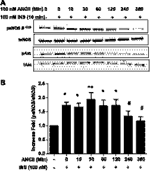
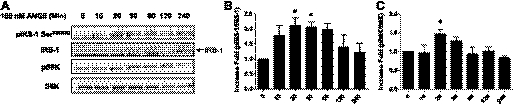
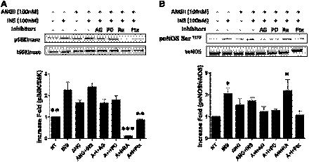
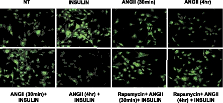
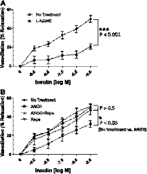
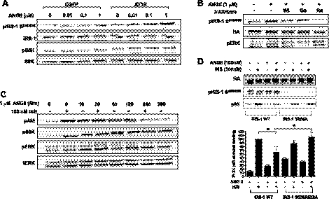
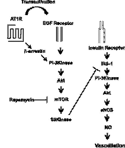
Elevated tissue levels of angiotensin II (ANG II) are associated with impairment of insulin actions in metabolic and cardiovascular tissues. ANG II-stimulated activation of mammalian target of rapamycin (mTOR)/p70 S6 kinase (p70S6K) in cardiovascular tissues is implicated in cardiac hypertrophy and vascular remodeling. However, the role of ANG II-stimulated mTOR/p70S6K in vascular endothelium is poorly understood. In the present study, we observed that ANG II stimulated p70S6K in bovine aortic endothelial cells. ANG II increased phosphorylation of insulin receptor substrate-1 (IRS-1) at Ser(636/639) and inhibited the insulin-stimulated phosphorylation of endothelial nitric oxide synthase (eNOS). An inhibitor of mTOR, rapamycin, attenuated the ANG II-stimulated phosphorylation of p70S6K and phosphorylation of IRS-1 (Ser(636/639)) and blocked the ability of ANG II to impair insulin-stimulated phosphorylation of eNOS, nitric oxide production, and mesenteric-arteriole vasodilation. Moreover, point mutations of IRS-1 at Ser(636/639) to Ala prevented the ANG II-mediated inhibition of insulin signaling. From these results, we conclude that activation of mTOR/p70S6K by ANG II in vascular endothelium may contribute to impairment of insulin-stimulated vasodilation through phosphorylation of IRS-1 at Ser(636/639). This ANG II-mediated impairment of vascular actions of insulin may help explain the role of ANG II as a link between insulin resistance and hypertension.


Figures

Fig. 1.
Fig. 1.
Angiotensin II (ANG II) inhibits insulin-stimulated phosphorylation of endothelial nitric oxide synathase (eNOS) and Akt. Bovine aortic endothelial cells (BAECs) were serum-starved overnight and then treated without or with ANG II (100 nmol/l) for the indicated times. Subsequently, cells were treated with insulin (100 nmol/l, 10 min) and harvested. The cell lysates were immunoblotted with antibodies against eNOS, Akt, phospho-eNOS (Ser1179), and phospho-Akt (Ser479). Pretreatment with ANG II inhibited the insulin-stimulated phosphorylation of eNOS and Akt. Pretreatment for 4 h and longer with ANG II inhibited insulin-stimulated phosphorylation of eNOS (means ± SE) *P ≤ 0.05; **P < 0.001; #P > 0.05 compared with no treatment, 1-way ANOVA with Dunnett's post hoc test.
Fig. 2.
Fig. 2.
ANG II alone stimulates phophorylation of insulin receptor substrate-1 (IRS-1) and 70-kDa ribosomal S6 kinase (p70S6K). BAECs were serum-starved overnight and then treated without or with ANG II (100 nmol/l) for the indicated periods. A: cells were harvested, and the cell lysates were immunoblotted with antibodies against IRS-1, phospho-IRS-1 (Ser636/639), p70S6K, and phospho-p70S6K (Thr389). B and C: treatment with ANG II stimulates phosphorylation of IRS-1 and p70S6K in a time-dependent manner. Three independent experiments were analyzed and shown in bar graphs (means ± SE). *P < 0.05 compared with no treatment, 1-way ANOVA with Dunnett's post hoc test.
Fig. 3.
Fig. 3.
Pretreatment with rapamycin restores the insulin-stimulated phophorylation of eNOS that is inhibited by ANG II. BAECs were serum-starved overnight and treated without or with AG1478 (5 μmol/l), PD-123319, (10 μM), rapamycin (25 nmol/l), or pertussis toxin (100 ng/ml) for 30 min prior to ANG II (100 nmol/l, 4 h). Then the cells were stimulated without or with insulin (100 nmol/l) for 10 min. Cells were harvested, and the cell lysates were immunoblotted with antibodies against phospho-(Thr389) and total p70S6K (A) and phospho- (Ser1179) and total eNOS (B). Immunoblots shown are representative of those that were independently repeated at least 3 times, and the quantifications of 3 independent experiments are shown as bar graphs (means ± SE). *P < 0.05; **P < 0.01; ***P < 0.001 compared with no treatment (A) and insulin treatment (B), 1-way ANOVA with Dunnettt's post hoc test.
Fig. 4.
Fig. 4.
Pretreatment with rapamycin restores the insulin-stimulated nitric oxide (NO) production that is inhibited by treatment with ANG II. BAECs were serum-starved overnight and treated without or with ANG II (100 nmol/l, 4 h). Then the cells were loaded with 4,5-diaminofluorescein diacetate, as described in materials and methods. Cells were then stimulated without or with insulin (100 nmol/l, 10 min). After insulin treatment, cells were fixed in 2% paraformaldehyde and visualized with an epifluorescent microscope, as described in materials and methods. Emission of green fluorescence is indicative of NO production. Experiments shown are representative of those that were repeated independently at least 3 times.
Fig. 5.
Fig. 5.
Pretreatment with rapamycin (Rapa) restores the insulin-stimulated vasodilation that is inhibited by treatment with ANG II. Mesenteric arterioles were isolated from Sprague-Dawley rats. The arterioles were cannulated onto glass micropipettes and pressurized to 70 mmHg using a Pressure Servo System and warmed to 37°C over 30 min for stabilization. The arterioles were treated without or with rapamycin (10 nmol/l, 30 min), and arterioles were treated without or with ANG II (100 nmol/l, 4 h). Arterioles were constricted with penylephrine (1 mmol/l) and then exposed to increasing concentrations of insulin. The vasodilatory activity of insulin on the arterioles was observed and recorded using a data acquisition system. In some experiments, NG-nitro-l-arginine methyl ester (l-NAME; 100 μmol/l, 30 min) was applied prior to insulin exposure. A: mesenteric arterioles were dilated in response to insulin in a concentration-dependent manner, and the insulin-stimulated dilation was inhibited by pretreatment of l-NAME. B: insulin-stimulated vasodilation was inhibited by ANG II and restored by pretreatment with rapamycin. No treatment (n = 6), ANG II (100 nmol/l for 4 h; n = 5), Rapa (10 nmol/l; n = 6), Rapa + Ang II (n = 6), and l-NAME (n = 11). %Vasorelaxation was calculated as described materials and methods. Means ± SE. *P < 0.05, ***P < 0.001; the data were analyzed by repeated measurements of 2-way ANOVA combined with Bonferroni post hoc test.
Fig. 6.
Fig. 6.
ANG II stimulates phosphorylation of IRS-1 at Ser636/639 and p70S6K in cells overexpressing ANG II receptor 1 (AT1R). NIH-3T3IR cells were transiently transfected with plasmid (pcDNA 3.1) containing AT1R cDNA. One day after transfection, the cells were serum-starved overnight and treated without or with indicated concentration of ANG II for 30 min. Cells were harvested, and the cell lysates were immunoblotted with antibodies as indicated. A: ANG II stimulated the phosphorylation of IRS-1 at Ser636/639 and p70S6K in the cells overexpressing AT1R. B: the ANG II-stimulated phosphorylation of IRS-1 at Ser636/639 was inhibited by wortmanin (100 nmol/l) or Rapa (25 nmol/l) but not by Go6976 (1 μmol/l). C: insulin (INS)-stimulated phosphorylation of Akt was attenuated by treatment with ANG II. D: point mutation of IRS-1 at Ser636/639A is protective from ANG II-induced inhibition of insulin-stimulated phosphorylation of Akt (means ± SE). *P < 0.05 compared with wild-type IRS-1, 2-way ANOVA with Bonferroni post hoc test.
Fig. 7.
Fig. 7.
Schematic diagram of ANG II signaling pathway in vascular endothelium. ANG II stimulates mammalian target of rapamycin (mTOR)/S6K through transactivation of epidermal growth factor (EGF) receptor activating mTOR/S6K that contributes to inhibition of insulin-stimulated phosphorylation of eNOS, production of NO, and vasodilation.

References

    1. Adochio R, Leitner JW, Hedlund R, Draznin B. Rescuing 3T3-L1 adipocytes from insulin resistance induced by stimulation of Akt-mammalian target of rapamycin/p70 S6 kinase (S6K1) pathway and serine phosphorylation of insulin receptor substrate-1: effect of reduced expression of p85alpha subunit of phosphatidylinositol 3-kinase and S6K1 kinase. Endocrinology 150: 1165–1173, 2009 - PubMed
    1. Ahn S, Kim J, Hara MR, Ren XR, Lefkowitz RJ. {beta}-Arrestin-2 Mediates Anti-apoptotic Signaling through Regulation of BAD Phosphorylation. J Biol Chem 284: 8855–8865, 2009 - PMC - PubMed
    1. Ali MS, Schieffer B, Delafontaine P, Bernstein KE, Ling BN, Marrero MB. Angiotensin II stimulates tyrosine phosphorylation and activation of insulin receptor substrate 1 and protein-tyrosine phosphatase 1D in vascular smooth muscle cells. J Biol Chem 272: 12373–12379, 1997 - PubMed
    1. Andreozzi F, Laratta E, Sciacqua A, Perticone F, Sesti G. Angiotensin II impairs the insulin signaling pathway promoting production of nitric oxide by inducing phosphorylation of insulin receptor substrate-1 on Ser312 and Ser616 in human umbilical vein endothelial cells. Circ Res 94: 1211–1218, 2004 - PubMed
    1. Arellano-Plancarte A, Hernandez-Aranda J, Catt KJ, Olivares-Reyes JA. Angiotensin-induced EGF receptor transactivation inhibits insulin signaling in C9 hepatic cells. Biochem Pharmacol 79: 733–745, 2010 - PMC - PubMed

Publication types

MeSH terms