. 2011 Oct 25;4(196):ra70.
doi: 10.1126/scisignal.2002278.

Regulation of insulin-like growth factor signaling by Yap governs cardiomyocyte proliferation and embryonic heart size

Affiliations

Regulation of insulin-like growth factor signaling by Yap governs cardiomyocyte proliferation and embryonic heart size

Mei Xin et al. Sci Signal. .

Abstract

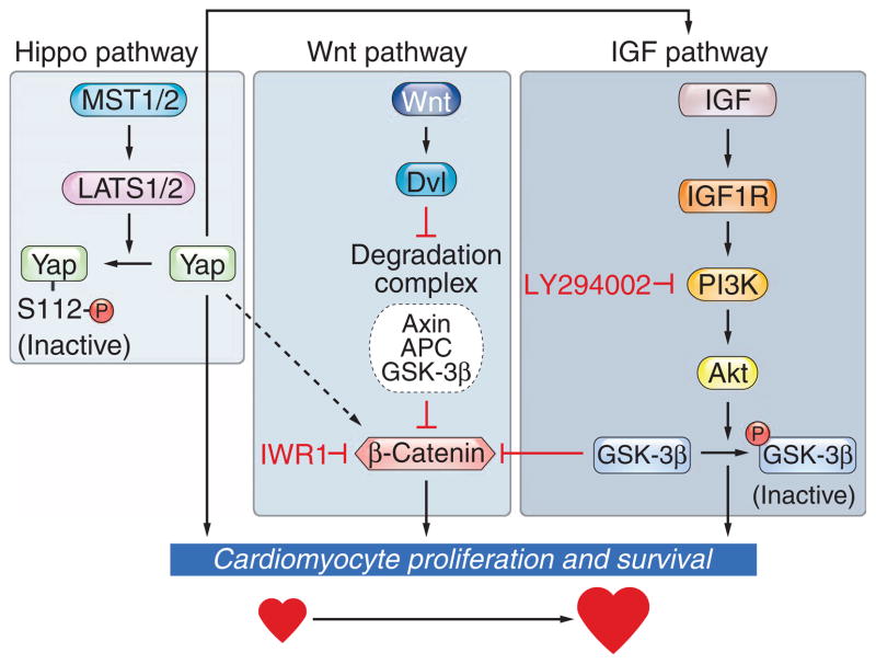
The Hippo signaling pathway regulates growth of the heart and other tissues. Hippo pathway kinases influence the activity of various targets, including the transcriptional coactivator Yap, but the specific role of Yap in heart growth has not been investigated. We show that Yap is necessary and sufficient for embryonic cardiac growth in mice. Deletion of Yap in the embryonic mouse heart impeded cardiomyocyte proliferation, causing myocardial hypoplasia and lethality at embryonic stage 10.5. Conversely, forced expression of a constitutively active form of Yap in the embryonic heart increased cardiomyocyte number and heart size. Yap activated the insulin-like growth factor (IGF) signaling pathway in cardiomyocytes, resulting in inactivation of glycogen synthase kinase 3β, which led to increased abundance of β-catenin, a positive regulator of cardiac growth. Our results point to Yap as a critical downstream effector of the Hippo pathway in the control of cardiomyocyte proliferation and a nexus for coupling the IGF, Wnt, and Hippo signaling pathways with the developmental program for heart growth.


Conflict of interest statement

Competing interests: The authors declare that they have no competing interests.

Figures

Fig. 1
Fig. 1
Yap is necessary and sufficient for embryonic heart growth. (A) Whole-mount, H&E, and phosphorylated H3 (PH3) immunostaining of YaploxP/+ (Control) and YaploxP/−; Nkx2.5-Cre (Yap nKO) embryos at E9.5. PH3 (red)–, Nkx2.5 (blue)–, and cardiac troponin T (green)–stained section of Yap nKO. lv, left ventricle; la, left atrium; v, common ventricle; a, atrium. n = 3 embryos per phenotype. Arrowheads, PH3-positive cells. Asterisks, blood cells. (B) Quantification of number of cardiomyocytes per section in control (Ctrl) and Yap nKO at E9.5, PH3 immunostaining in Ctrl and Yap nKO at E9.5, and of PH3-positive cells in wild-type (WT) and βMHC-YapS112A transgenic (Tg) mice at E10.5. n = 3 embryos per genotype. Data are presented as means ± SEM. *P < 0.05, **P < 0.01, by t test. (C) H&E-stained sections of embryonic hearts from βMHC-YapS112A Tg and WT mice at E14.5. The top panels show the entire heart, and the lower panels show higher magnification of the ventricles of the hearts. la, left atrium; lv, left ventricle; ra, right atrium; rv, right ventricle. Scale bars, 100 μm. (D) H&E staining of transverse section of WT and βMHC-YapS112A Tg embryos at E10.5 (left column). PH3-stained sections of hearts from WT and βMHC-YapS112A Tg embryos at E10.5 (middle column). Higher magnification of H&E staining of the left ventricles (right column). la, left atrium; lv, left ventricle; ra, right atrium; rv, right ventricle. Scale bars, 100 μm.
Fig. 2
Fig. 2
Yap promotes neonatal cardiomyocyte proliferation in vitro. (A) BrdU (red) and α-actinin (green) staining of neonatal rat cardiomyocytes infected with adenovirus expressing GFP or YapS112A. Arrowheads, BrdU-positive cells. Scale bar, 20 μm. (B) Quantification of BrdU incorporation assays. Cells were counted in six randomly selected fields for each condition. Number of cells counted per field was >100. Data are presented as means ± SEM. ***P < 0.001 by t test. (C) Quantification of PH3-positive cardiomyocytes. Cells were counted in eight randomly selected fields for each condition. Number of cells counted per field was >100. Data are presented as means ± SEM. ***P < 0.001 by t test. (D) Representative image of cardiomyocytes infected with Ad-YapS112A, immunostained for Aurora B kinase (red) and α-actinin (green). Arrows, cells undergoing cytokinesis. Scale bar, 20 μm. (E) Quantification of Aurora B kinase immunostaining. Cells were counted in six randomly selected fields for each condition. Number of cells counted per field was >100. Data are presented as means ± SEM. **P < 0.01 by t test. (F) Quantification of cardiomyocyte number by cardiac α-actinin staining. Cells were counted in nine randomly selected fields for each condition. Number of cells counted per field was >100. Data are presented as means ± SEM. ***P < 0.001 by ANOVA with the Bonferroni post test.
Fig. 3
Fig. 3
Yap increases the activity of the IGF signaling pathway. (A) Neonatal rat cardiomyocytes were infected with adenovirus expressing GFP or YapS112A and immunoblotted with antibodies against the indicated proteins. Glyceraldehyde-3-phosphate dehydrogenase (GAPDH) was used as loading control. (B) Quantification of abundance of IGF, Akt, and β-catenin signaling proteins in neonatal rat cardiomyocytes infected with adenoviruses expressing GFP or YapS112A. Values were determined with data from four Western blots. Protein quantification was normalized to Ad-GFP–infected cells. *P < 0.05, Mann-Whitney test. (C) Neonatal rat cardiomyocytes were infected with adenovirus expressing the indicated genes followed by transfection with a TOPflash luciferase reporter. Luciferase activity was normalized to β-galactosidase activity. Data are presented as means ± SEM. *P < 0.05, Mann-Whitney test. (D) Aurora B kinase immunostaining was quantified after cardiomyocytes were infected with the indicated recombinant adenovirus and incubated with the β-catenin inhibitor IWR1. Data are presented as means ± SEM. ***P < 0.001, Mann-Whitney test. (E) Neonatal rat cardiomyocytes were infected with adenovirus expressing GFP or YapS112A, serum-starved, stimulated with IGF, and immunoblotted for active Akt. GAPDH was used as loading control. Protein quantification was normalized to Ad-GFP without IGF treatment. Quantification of three Western blots is shown. *P < 0.05, ***P < 0.001 by ANOVA with the Bonferroni post test. (F) Neonatal rat cardiomyocytes were infected with recombinant adeno-virus expressing GFP or YapS112A, transfected with TOPflash luciferase reporter, and incubated with the PI3K inhibitor LY294002. Luciferase activity was normalized to β-galactosidase activity. Data are presented as means ± SEM. **P < 0.01, Mann-Whitney test. (G) Neonatal rat cardiomyocytes were infected with adenovirus expressing GFP or YapS112A, incubated with LY294002, and immunoblotted for active β-catenin. Quantification of protein expression from three Western blots is shown in the bar graph, with a representative Western blot shown below. **P < 0.01, ANOVA with the Bonferroni post test. (H) Neonatal rat cardiomyocytes were infected with adenovirus expressing GFP or YapS112A, transfected with small interfering RNAs (siRNAs) against β-catenin or IGF1R, and immunoblotted for β-catenin or IGF1R antibodies to confirm knockdown. (I) Quantification of Aurora B–positive cells. Cells were counted in eight randomly selected fields for each condition. Number of cells counted per field was >100. Data are presented as means ± SEM. ***P < 0.001, ANOVA with the Bonferroni post test.
Fig. 4
Fig. 4
Model showing a role for Yap in governing cardiac growth and survival by interlinking the Hippo, IGF, and Wnt pathways. Yap promotes cardiomyocyte proliferation through activation of IGF signaling and stabilization of the active form of β-catenin. Inhibition of IGF signaling or β-catenin blocks Yap-induced cardiomyocyte proliferation. Yap interlinks the Hippo pathway (MST1/2 and LATS1/2), the Wnt pathway [Wnt, Disheveled (Dvl), Axin, APC, and GSK-3β], and the IGF pathway (IGF, IGF1R, PI3K, Akt, and GSK-3β) to control β-catenin signaling and regulate cardiac growth and development. Each of the three pathways influenced by Yap is demarcated by a box.

References

    1. Vincent SD, Buckingham ME. How to make a heart: The origin and regulation of cardiac progenitor cells. Curr Top Dev Biol. 2010;90:1–41. - PubMed
    1. Pan D. The hippo signaling pathway in development and cancer. Dev Cell. 2010;19:491–505. - PMC - PubMed
    1. Huang J, Wu S, Barrera J, Matthews K, Pan D. The Hippo signaling pathway coordinately regulates cell proliferation and apoptosis by inactivating Yorkie, the Drosophila homolog of YAP. Cell. 2005;122:421–434. - PubMed
    1. Zhang H, Pasolli HA, Fuchs E. Yes-associated protein (YAP) transcriptional coactivator functions in balancing growth and differentiation in skin. Proc Natl Acad Sci USA. 2011;108:2270–2275. - PMC - PubMed
    1. Schlegelmilch K, Mohseni M, Kirak O, Pruszak J, Rodriguez JR, Zhou D, Kreger BT, Vasioukhin V, Avruch J, Brummelkamp TR, Camargo FD. Yap1 acts downstream of α-catenin to control epidermal proliferation. Cell. 2011;144:782–795. - PMC - PubMed

Publication types

MeSH terms