. 2011 Oct;13(10):912-22.
doi: 10.1593/neo.11576.

Elucidating the mechanism of regulation of transforming growth factor β Type II receptor expression in human lung cancer cell lines

Affiliations

Elucidating the mechanism of regulation of transforming growth factor β Type II receptor expression in human lung cancer cell lines

Sunil K Halder et al. Neoplasia. 2011 Oct.

Abstract

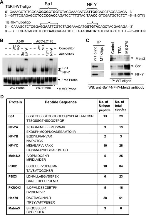
Lung carcinogenesis in humans involves an accumulation of genetic and epigenetic changes that lead to alterations in normal lung epithelium, to in situ carcinoma, and finally to invasive and metastatic cancers. The loss of transforming growth factor β (TGF-β)-induced tumor suppressor function in tumors plays a pivotal role in this process, and our previous studies have shown that resistance to TGF-β in lung cancers occurs mostly through the loss of TGF-β type II receptor expression (TβRII). However, little is known about the mechanism of down-regulation of TβRII and how histone deacetylase (HDAC) inhibitors (HDIs) can restore TGF-β-induced tumor suppressor function. Here we show that HDIs restore TβRII expression and that DNA hypermethylation has no effect on TβRII promoter activity in lung cancer cell lines. TGF-β-induced tumor suppressor function is restored by HDIs in lung cancer cell lines that lack TβRII expression. Activation of mitogen-activated protein kinase/extracellular signal-regulated kinase pathway by either activated Ras or epidermal growth factor signaling is involved in the down-regulation of TβRII through histone deacetylation. We have immunoprecipitated the protein complexes by biotinylated oligonucleotides corresponding to the HDI-responsive element in the TβRII promoter (-127/-75) and identified the proteins/factors using proteomics studies. The transcriptional repressor Meis1/2 is involved in repressing the TβRII promoter activity, possibly through its recruitment by Sp1 and NF-YA to the promoter. These results suggest a mechanism for the downregulation of TβRII in lung cancer and that TGF-β tumor suppressor functions may be restored by HDIs in lung cancer patients with the loss of TβRII expression.


Figures

Figure 1
Figure 1
Restoration of TGF-β-induced tumor suppressor functions by TSA. (A) VMRC-LCD cells were transiently cotransfected with CMV-β-gal, p3TP-Lux or (CAGA)9 MLP-Luc plasmids, together with the indicated expression plasmids. Cells were treated with TGF-β alone or both TGF-β and TSA. Normalized luciferase activity was expressed as the mean ± SD of triplicate measurements. *P < .05 compared with the corresponding control. **P < .05 when compared between the two data points. ***P < .05 when compared with the corresponding control. (B) Thymidine incorporation assay: A549 and VMRC-LCD cells were treated with increasing amounts of TSA (left). VMRC-LCD cells were also treated with increasing doses of TSA in the presence or absence of SB-431542 (SB) (right). Radioactivity incorporated without TSA treatment is considered as 100%, and the results are expressed as the mean ± SD. *P < .05 compared with the corresponding control. ΔP < .05 and ΔΔP < .05 compared with the corresponding SB treatment. (C) Soft agarose assay was performed as described in Materials and Methods. In addition, 5 x 10³ ACC-LC-176 cells were plated on soft agar as described above except that TSA and/or SB-431542 were added on the top agar layer every third day. Each data point represents the number of colonies from an average of three values. *P < .05 compared with the corresponding control. **P < .05 when compared between the two data points. (D) In vivo tumorigenicity assay: ACC-LC-176, VMRC-LCD, and A549 were injected subcutaneously behind the anterior fore limb of BALB/c athymic nude mice. TSA (25 µg/d per mice) was administered intraperitoneally. Growth curves are plotted from the mean volume ± SD of tumors from six mice in each group. *P < .05 compared with the corresponding control.
Figure 2
Figure 2
The HDIs restore TβRII expression in VMRC-LCD (A) and ACC-LC-176 (B) cells but not in A549 (C) cells. Human lung tumor cell lines, VMRC-LCD, ACC-LC-176 (without TβRII expression), and A549 (with TβRII expression), were treated with HDIs and AZA. RT-PCR analyses for TβRII gene expression were performed with total RNA as described previously [5].
Figure 3
Figure 3
TSA treatment restores Ras- and EGF-mediated down-regulation of TβRII expression. (A) Lysates from RIE-iRas cells treated with 5 mM IPTG were analyzed by Western blots using anti-TβRII and anti-Pan-Ras antibodies. (B) Lysates from RIE-iRas cells treated with IPTG in the presence or absence of TSA for indicated times were analyzed by Western blots using anti-TβRII, anti-Ac-histone-H3, anti-Ac-histone-H4, and anti-β-actin antibodies. (C) Lysates from RIE-iRas cells treated with IPTG in the presence of TSA, PD98059, or U0126 for 24 and 48 hours were analyzed by Western blot analyses. (D) Lysates from parental RIE cells treated with EGF (10 ng/ml) in the presence of TSA or U0126 for 24 and 48 hours were analyzed by Western blots.
Figure 4
Figure 4
The promoter region of TβRII (-127/-75) is required for its induction by MS-275 and TSA. (A) A schematic diagram representing the multiple regulatory elements within the TβRII promoter. The TβRII promoter luciferase constructs and β-gal plasmid were transiently cotransfected into ACC-LC-176 cells. Transfected cells were treated with TSA, MS-275, and/or AZA (B). Luciferase activity was normalized and expressed as the mean ± SD. *P < .05 and **P < .05 compared with the corresponding control.
Figure 5
Figure 5
Identification of proteins that bind to HRE of TβRII promoter. (A) The WT and mutated (Sp1 and NF-YA sites mutated) oligonucleotide sequences containing HRE in the TβRII promoter are shown. (B) An EMSA was performed using 32P-labeled WT oligo (-115/-64 of TβRII promoter, WO) and mutant oligo probes by incubating nuclear lysates from A549 and ACC-LC-176 cells. In competitive binding or supershift assays, unlabeled WO or MO or antibodies against Sp1 or NF-Y was preincubated with the nuclear extract. DNA-protein complexes were resolved in a 5% native acrylamide gel. (C) Biotinylated double-stranded oligo corresponding to the -127/-65 sequence (WT oligo) or mutant oligo (MT oligo) was mixed with equal amounts of nuclear extracts prepared from ACC-LC-176 cells treated with TSA or MS-275. DNA-protein complexes were precipitated with streptavidin-agarose beads and subjected to Western blot analyses for Sp1, NF-YA, and Meis2. (D) DAPA was performed using WT and mutant oligos for proteomics studies. DNA-protein complexes were precipitated and resolved in 10% SDS-PAGE. Proteins were eluted, digested with trypsin or chymotrypsin, and finally analyzed by LC-MS/MS. The number of unique peptides and the number of total spectra including two representative peptide sequences for each protein detected by MS are shown.
Figure 6
Figure 6
Meis2 inhibits HDI-induced TβRII promoter activity. (A) Vector control and Meis2-overexpressing ACC-LC-176 clones were transiently cotransfected with CMV-β-gal and TβRII promoter reporter plasmids. Transfected cells were treated with MS-275, and the normalized relative luciferase activities are shown. *P < .05 compared with the corresponding control. **P < .05 compared between corresponding treatment points. (B) ACC-LC-176 cells were transiently cotransfected with CMV-β-gal and various plasmid constructs that contain WT HRE, HRE (Sp1 mutant), HRE (NF-YA mutant), or HRE (double mutant). Transfected cells were treated with MS-275, and the relative luciferase activities are shown. *P < .05 compared with the corresponding control. **P < .05 compared with the corresponding WT treatment point. (C) ACC-LC-176 cells were transiently cotransfected with CMV-β-gal and various plasmid constructs as described above. Transfected cells were treated with MS-275 with or without EGF (10 ng/ml), and the relative luciferase activities are shown. *P < .05 compared with the corresponding control. **P < .05 compared with corresponding treatment points. ΔP < .05 when compared with the WT treatment point. (D) Meis2 physically interacts with Sp1 and NF-YA. 293T cells were cotransfected with Meis2-HA and Sp1-Flag (left) or Meis2-Flag and NF-YA-HA (right panel) expression vectors. Cell lysates were subjected to immunoprecipitation (IP) with anti-Flag or anti-HA antibodies, and the presence of Meis2 in the complex was detected by Western blot analysis. Expressions of Meis2-HA, Meis2-Flag, Sp1-Flag, and NF-YA-HA proteins were determined (bottom panels). (E) Hypothetical model for the loss of TGF-β tumor suppressor function in lung cancer. Up-regulation/activation of EGF receptor (EGFR) and/or oncogenic activation of Ras in lung cancer result in the activation of MEK/ERK pathway that leads to down-regulation of TβRII. MEK1/2 inhibitors block the down-regulation of TβRII induced by activated Ras or EGFR. The reduced level of TβRII is due to the recruitment of HDAC activity on the promoter by transcription factors (TF) with subsequent replacement of histone acetyl transferase proteins including p300 and CBP. TGF-β-induced tumor suppressor function may be restored by treatment with HDIs in lung tumors that are resistant to TGF-β due to the loss of TβRII expression.

References

    1. Nagatake M, Takagi Y, Osada H, Uchida K, Mitsudomi T, Saji S, Shimokata K, Takahashi T, Takahashi T. Somatic in vivo alterations of the DPC4 gene at 18q21 in human lung cancers. Cancer Res. 1996;56:2718–2720. - PubMed
    1. Uchida K, Nagatake M, Osada H, Yatabe Y, Kondo M, Mitsudomi T, Masuda A, Takahashi T, Takahashi T. Somatic in vivo alterations of the JV18-1 gene at 18q21 in human lung cancers. Cancer Res. 1996;56:5583–5585. - PubMed
    1. Osada H, Tatematsu Y, Masuda A, Saito T, Sugiyama M, Yanagisawa K, Takahashi T. Heterogeneous transforming growth factor (TGF)-β unresponsiveness and loss of TGF-β receptor type II expression caused by histone deacetylation in lung cancer cell lines. Cancer Res. 2001;61:8331–8339. - PubMed
    1. Hougaard S, Norgaard P, Abrahamsen N, Moses HL, Spang-Thomsen M, Skovgaard Poulsen H. Inactivation of the transforming growth factor β type II receptor in human small cell lung cancer cell lines. Br J Cancer. 1999;79:1005–1011. - PMC - PubMed
    1. Anumanthan G, Halder SK, Osada H, Takahashi T, Massion PP, Carbone DP, Datta PK. Restoration of TGF-β signalling reduces tumorigenicity in human lung cancer cells. Br J Cancer. 2005;93:1157–1167. - PMC - PubMed

Publication types

MeSH terms

LinkOut - more resources