

Ectopic overexpression of Sonic Hedgehog (Shh) induces stromal expansion and metaplasia in the adult murine pancreas
- PMID: 22028618
- PMCID: PMC3201569
- DOI: 10.1593/neo.11088
Ectopic overexpression of Sonic Hedgehog (Shh) induces stromal expansion and metaplasia in the adult murine pancreas
Abstract
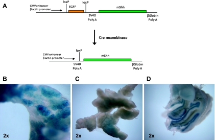
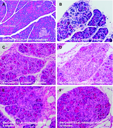
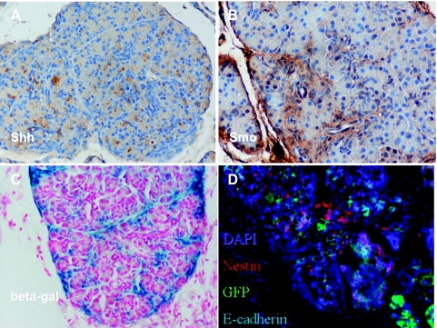
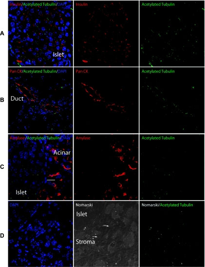
Ligand-dependent activation of the Hedgehog (Hh) signaling pathway has been implicated in both tumor initiation and metastasis of pancreatic ductal adenocarcinoma (PDAC). Prior studies in genetically engineered mouse models (GEMMs) have assessed the role of Hh signaling by cell autonomous expression of a constitutively active Gli2 within epithelial cells. On the contrary, aberrant pathway reactivation in the human exocrine pancreas occurs principally as a consequence of Sonic Hh ligand (Shh) overexpression from epithelial cells. To recapitulate the cognate pathophysiology of Hh signaling observed in the human pancreas, we examined GEMM where Hh ligand is conditionally overexpressed within the mature exocrine pancreas using a tamoxifen-inducible Elastase-Cre promoter (Ela-CreERT2;LSL-mShh). We also facilitated potential cell autonomous epithelial responsiveness to secreted Hh ligand by generating compound transgenic mice with concomitant expression of the Hh receptor Smoothened (Ela-CreERT2;LSL-mShh;LSL-mSmo). Of interest, none of these mice developed intraductal precursor lesions or PDAC during the follow-up period of up to 12 months after tamoxifen induction. Instead, all animals demonstrated marked expansion of stromal cells, consistent with the previously described epithelial-to-stromal paracrine Hh signaling. Hh responsiveness was mirrored by the expression of primary cilia within the expanded mesenchymal compartment and the absence within mature acinar cells. In the absence of cooperating mutations, Hh ligand overexpression in the mature exocrine pancreas is insufficient to induce neoplasia, even when epithelial cells coexpress the Smo receptor. This autochthonous model serves as a platform for studying epithelial stromal interactions in pancreatic carcinogenesis.
Figures
Similar articles
-
Primary cilia regulate Gli/Hedgehog activation in pancreas.Proc Natl Acad Sci U S A. 2010 Jun 1;107(22):10109-14. doi: 10.1073/pnas.0909900107. Epub 2010 May 17. Proc Natl Acad Sci U S A. 2010. PMID: 20479231 Free PMC article.
-
Hedgehog signaling is required for effective regeneration of exocrine pancreas.Gastroenterology. 2008 Aug;135(2):621-31. doi: 10.1053/j.gastro.2008.04.011. Epub 2008 Apr 16. Gastroenterology. 2008. PMID: 18515092 Free PMC article.
-
Aberrant Hedgehog ligands induce progressive pancreatic fibrosis by paracrine activation of myofibroblasts and ductular cells in transgenic zebrafish.PLoS One. 2011;6(12):e27941. doi: 10.1371/journal.pone.0027941. Epub 2011 Dec 2. PLoS One. 2011. PMID: 22164219 Free PMC article.
-
Paracrine sonic hedgehog signaling derived from tumor epithelial cells: a key regulator in the pancreatic tumor microenvironment.Crit Rev Eukaryot Gene Expr. 2012;22(2):97-108. doi: 10.1615/critreveukargeneexpr.v22.i2.20. Crit Rev Eukaryot Gene Expr. 2012. PMID: 22856428 Review.
-
Sonic Hedgehog in pancreatic cancer: from bench to bedside, then back to the bench.Surgery. 2012 Sep;152(3 Suppl 1):S19-32. doi: 10.1016/j.surg.2012.05.030. Epub 2012 Jul 6. Surgery. 2012. PMID: 22770959 Free PMC article. Review.
Cited by
-
Inflammatory and Senescent Phenotype of Pancreatic Stellate Cells Induced by Sqstm1 Downregulation Facilitates Pancreatic Cancer Progression.Int J Biol Sci. 2019 Apr 21;15(5):1020-1029. doi: 10.7150/ijbs.27825. eCollection 2019. Int J Biol Sci. 2019. PMID: 31182922 Free PMC article.
-
A Quick Guide to CAF Subtypes in Pancreatic Cancer.Cancers (Basel). 2023 May 4;15(9):2614. doi: 10.3390/cancers15092614. Cancers (Basel). 2023. PMID: 37174079 Free PMC article. Review.
-
Overcoming intratumor heterogeneity of polygenic cancer drug resistance with improved biomarker integration.Neoplasia. 2012 Dec;14(12):1278-89. doi: 10.1593/neo.122096. Neoplasia. 2012. PMID: 23308059 Free PMC article.
-
Oncogenic KRAS Regulates Tumor Cell Signaling via Stromal Reciprocation.Cell. 2016 May 5;165(4):910-20. doi: 10.1016/j.cell.2016.03.029. Epub 2016 Apr 14. Cell. 2016. PMID: 27087446 Free PMC article.
-
Mechanistic insights into self-reinforcing processes driving abnormal histogenesis during the development of pancreatic cancer.Am J Pathol. 2013 Apr;182(4):1078-86. doi: 10.1016/j.ajpath.2012.12.004. Epub 2013 Jan 31. Am J Pathol. 2013. PMID: 23375449 Free PMC article. Review.
References
-
- Jemal A, Siegel R, Xu J, Ward E. Cancer statistics, 2010. CA Cancer J Clin. 2010;60:277–300. - PubMed
-
- Berman DM, Karhadkar SS, Maitra A, Montes De Oca R, Gerstenblith MR, Briggs K, Parker AR, Shimada Y, Eshleman JR, Watkins DN, et al. Widespread requirement for Hedgehog ligand stimulation in growth of digestive tract tumours. Nature. 2003;425:846–851. - PubMed
Publication types
MeSH terms
Substances
Grants and funding
LinkOut - more resources
Full Text Sources
Miscellaneous