. 2011 Oct;9(10):e1001176.
doi: 10.1371/journal.pbio.1001176. Epub 2011 Oct 18.

Mouse PRDM9 DNA-binding specificity determines sites of histone H3 lysine 4 trimethylation for initiation of meiotic recombination

Affiliations

Mouse PRDM9 DNA-binding specificity determines sites of histone H3 lysine 4 trimethylation for initiation of meiotic recombination

Corinne Grey et al. PLoS Biol. 2011 Oct.

Abstract

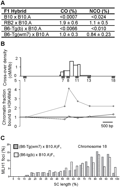
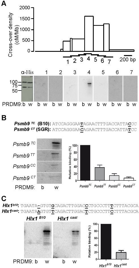
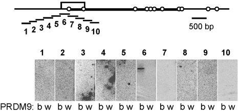
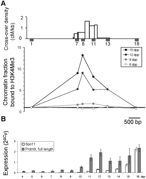
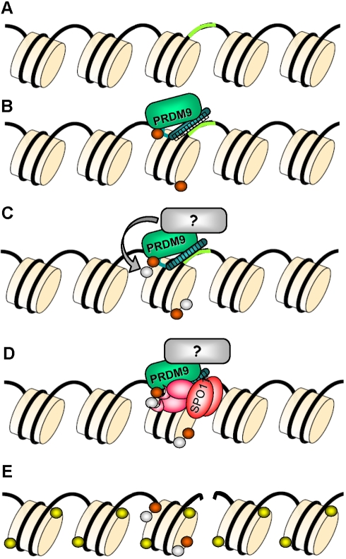
Meiotic recombination generates reciprocal exchanges between homologous chromosomes (also called crossovers, COs) that are essential for proper chromosome segregation during meiosis and are a major source of genome diversity by generating new allele combinations. COs have two striking properties: they occur at specific sites, called hotspots, and these sites evolve rapidly. In mammals, the Prdm9 gene, which encodes a meiosis-specific histone H3 methyltransferase, has recently been identified as a determinant of CO hotspots. Here, using transgenic mice, we show that the sole modification of PRDM9 zinc fingers leads to changes in hotspot activity, histone H3 lysine 4 trimethylation (H3K4me3) levels, and chromosome-wide distribution of COs. We further demonstrate by an in vitro assay that the PRDM9 variant associated with hotspot activity binds specifically to DNA sequences located at the center of the three hotspots tested. Remarkably, we show that mutations in cis located at hotspot centers and associated with a decrease of hotspot activity affect PRDM9 binding. Taken together, these results provide the direct demonstration that Prdm9 is a master regulator of hotspot localization through the DNA binding specificity of its zinc finger array and that binding of PRDM9 at hotspots promotes local H3K4me3 enrichment.


Conflict of interest statement

The authors have declared that no competing interests exist.

Figures

Figure 1
Figure 1. Influence of the PRDM9 zinc fingers on meiotic recombination and H3K4 trimethylation at recombination hotspots.
(A) Recombination activity at the Psmb9 hotspot is controlled by the PRDM9 zinc finger array. COs and NCOs at site “38” were measured by sperm typing . The 95% confidence intervals for recombinant product frequencies are calculated as described in . The difference in CO frequency between RB2×B10.A and B6-Tg(wm7)xB10.A is marginally significant (p = 0.03, two-sided heteroscedastic Student's t test). Values for B10×B10.A are from and for RB2×B10.A from . (B) H3K4me3 enrichment at the Psmb9 hotspot is controlled by the PRDM9 zinc finger array. Top panel, distribution of COs and positions of STSs used for chromatin analysis along the Psmb9 hotspot, from . The chromatin fraction bound to H3K4me3, normalized to the sequence-tagged site (STS) Psmb9-1 (STS1, the 5′ most flanking STS), was determined for each STS, as described in . Open circles, (B6-Tg(b)×B10.A)F1; grey circles, (B6-Tg(wm7)×B6)F1. The statistical analysis is shown in Table S2. (C) CO chromosome-wide distribution is controlled by the PRDM9 zinc finger array. The distribution of MLH1 foci along Chromosome 18 was determined as described in in pachytene chromosome spreads from (B6-Tg(b)×B10.A)F1 (spotted columns) and (B6-Tg(wm7)×B10.A)F1 (grey columns) hybrids. Each column represents the percentage of MLH1 foci per 5% interval of SC length. According to the size of chromosome 18 (90.722.031 bp, NCBI m37), one interval corresponds to about 4.5 Mb. Results for the two hybrids are comparable to the CO distribution observed in (B10×B10.A)F1 and (RB2×B10.A)F1 hybrids, respectively, but are significantly different from each other (Figure S3 and Table S4).
Figure 2
Figure 2. PRDM9 binds in vitro to meiotic recombination hotspots.
(A) Detection by southwestern blotting of PRDM9 binding at the Psmb9 recombination hotspot. Upper panel, CO distribution along the Psmb9 hotspot . Horizontal bars show the positions of the DNA probes (numbered from 1 to 7) used for southwestern experiments. Lower panel, PRDM9 (b, His-PRDM9b; w, His-PRDM9wm7) was probed with anti-His antibody and the radio-labeled double-stranded DNA probes 1–7 (about 200 bp). The molecular weights of His-PRDM9b and His-PRDM9wm7 are 101 kDa and 98 kDa, respectively. The bands with lower molecular weights correspond to PRDM9 degradation products. (B) Effect of SNPs at the center of the Psmb9 hotspot on the in vitro binding of PRDM9. The sequence of the likely PRDM9wm7 binding sequence is shown, and the SNPs between the B10 and B10.MOL-SGR strains are underlined (see FigS5 for in silico prediction). PRDM9b and PRDM9wm7 were probed with radio-labeled double-stranded oligonucleotides that carried the four possible SNP combinations (60 bp, Table S14). The amount of signal due to binding of each probe to PRDM9wm7 is shown (with standard error), relative to Psmb9TC. The decrease of binding to the double mutant probe Psmb9 CT (0.07% binding relative to Psmb9 TC) is consistent with a cumulative effect of each single mutant (0.36% and 0.14% binding relative to Psmb9 TC) suggesting their effects are independent. (C) Analysis by southwestern blotting of PRDM9 binding to the putative PRDM9wm7 binding motif at the center of the Hlx1 hotspot . The likely PRDM9wm7 binding sequences in the B10 and CAST strains are shown, with SNPs underlined (see Figure S5 for in silico prediction). PRDM9b and PRDM9wm7 were probed with radio-labeled double-stranded oligonucleotides that carried B10 or CAST allele (41 bp, Table S14). Signal intensities of the binding of the Hlx1B10 and Hlx1cast probes (relative to HlxB10) to PRDM9wm7 are shown.
Figure 3
Figure 3. PRDM9 binds to the G7c recombination hotspot.
Top, map of the genomic region of the G7c hotspot, located in the seventh intron of the D6S56E-3 gene on Chromosome 17. The open box represents the 800 bp interval with the highest density of exchanges, as mapped in . The open circles indicate the positions of the SNPs that are polymorphic in the hybrids used for measuring recombination (G7cb/a, see Table S7). The interval drawn as a thick line is the interval amplified by allele-specific PCR for measuring the recombination frequencies shown on Table S7. The position of the 10 probes used for southwestern blotting is shown underneath. Bottom, PRDM9 (b, His-PRDM9b; w, His-PRDM9wm7) was probed with the radio-labeled double-stranded DNA probes 1–10 (about 250 bp).
Figure 4
Figure 4. Kinetics of H3K4 trimethylation and Prdm9 expression in testes of prepuberal mice.
(A) Top panel, distribution of COs and positions of STSs used for chromatin analysis along the Psmb9 hotspot, from . The fraction of chromatin bound to H3K4me3, normalized to STS1 (the 5′ most flanking STS), was determined along the Psmb9 hotspot in whole testes from prepuberal R209 mice, as described previously . Data in 9 dpp mice are from . (B) Steady-state levels of Spo11 (white) and Prdm9 (full length, gray) transcripts were determined in whole testes from 4 to 18 dpp R209 mice. Given that the first wave of entry into meiosis is relatively synchronous, the decrease in Prdm9 transcript levels at 13 and 14 dpp may indicate a transient expression of Prdm9 at the beginning of meiotic prophase (10–12 dpp). The significant increase detected later at 18 dpp parallels the second wave of entry into meiosis.
Figure 5
Figure 5. Model of hotspot specification by PRDM9.
(A) The DNA and several nucleosomes are represented. A DNA sequence motif recognized by PRDM9 is represented in green. (B) PRDM9 binds to its target DNA motif through the zinc finger array and catalyzes H3K4me3 (orange). (C) A protein partner of PRDM9 may catalyze another post-translational histone modification (grey), allowing for the formation of a hotspot-specific signature. (D) PRDM9, a partner, or other component of the chromatin may recruit the recombination initiation complex containing SPO11 or may create a favorable chromatin environment allowing access of SPO11 to the DNA. (E) A DSB is formed by SPO11 and triggers the phosphorylation of histone H2Ax (yellow) in the surrounding nucleosomes. The DSB is then repaired by homologous recombination and lead to a CO or to gene conversion without CO.

References

    1. Arnheim N, Calabrese P, Tiemann-Boege I. Mammalian meiotic recombination hot spots. Annu Rev Genet. 2007;41:369–399. - PubMed
    1. Buard J, de Massy B. Playing hide and seek with mammalian meiotic crossover hotspots. Trends Genet. 2007;23:301–309. - PubMed
    1. Paigen K, Petkov P. Mammalian recombination hot spots: properties, control and evolution. Nat Rev Genet. 2010;11:221–233. - PMC - PubMed
    1. Jeffreys A. J, Kauppi L, Neumann R. Intensely punctate meiotic recombination in the class II region of the major histocompatibility complex. Nat Genet. 2001;29:217–222. - PubMed
    1. Jeffreys A. J, Neumann R, Panayi M, Myers S, Donnelly P. Human recombination hot spots hidden in regions of strong marker association. Nat Genet. 2005;37:601–606. - PubMed

Publication types