. 2011 Oct;7(10):e1002317.
doi: 10.1371/journal.pgen.1002317. Epub 2011 Oct 20.

Identification of widespread ultra-edited human RNAs

Affiliations

Identification of widespread ultra-edited human RNAs

Shai Carmi et al. PLoS Genet. 2011 Oct.

Abstract

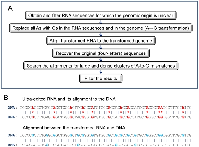
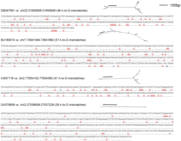
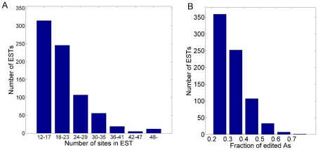
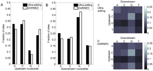
Adenosine-to-inosine modification of RNA molecules (A-to-I RNA editing) is an important mechanism that increases transciptome diversity. It occurs when a genomically encoded adenosine (A) is converted to an inosine (I) by ADAR proteins. Sequencing reactions read inosine as guanosine (G); therefore, current methods to detect A-to-I editing sites align RNA sequences to their corresponding DNA regions and identify A-to-G mismatches. However, such methods perform poorly on RNAs that underwent extensive editing ("ultra"-editing), as the large number of mismatches obscures the genomic origin of these RNAs. Therefore, only a few anecdotal ultra-edited RNAs have been discovered so far. Here we introduce and apply a novel computational method to identify ultra-edited RNAs. We detected 760 ESTs containing 15,646 editing sites (more than 20 sites per EST, on average), of which 13,668 are novel. Ultra-edited RNAs exhibit the known sequence motif of ADARs and tend to localize in sense strand Alu elements. Compared to sites of mild editing, ultra-editing occurs primarily in Alu-rich regions, where potential base pairing with neighboring, inverted Alus creates particularly long double-stranded RNA structures. Ultra-editing sites are underrepresented in old Alu subfamilies, tend to be non-conserved, and avoid exons, suggesting that ultra-editing is usually deleterious. A possible biological function of ultra-editing could be mediated by non-canonical splicing and cleavage of the RNA near the editing sites.


Conflict of interest statement

The authors have declared that no competing interests exist.

Figures

Figure 1
Figure 1. The computational procedure used to detect ultra-edited RNA.
(A) An outline of the procedure. (B) An illustration of the transformation algorithm. Top panel: an alignment between (hypothetical) ultra-edited RNA and its DNA source. A-to-G mismatches are denoted with red stars and mismatching nucleotides are highlighted in red. Bottom panel: alignment of the same sequences, but where each A was transformed to G (in both the DNA and the RNA). Transformed nucleotides are highlighted in light blue. A-to-G mismatches, but also A-A matches, become G-G matches in the transformed sequences. The transformed DNA and RNA therefore perfectly align.
Figure 2
Figure 2. The alignment of typical ultra-edited RNAs to the genome.
The alignments were generated using NCBI BLAST (http://www.ncbi.nlm.nih.gov/BLAST/Blast.cgi). The RNA secondary structure (RNAFold [48]) is also shown. The bar indicates approximately 100 base pairs. All ESTs display tens of A-to-G mismatches as well as a clear dsRNA structure.
Figure 3
Figure 3. The number of editing sites and the editing rate in the ultra-edited ESTs.
(A) The number of ESTs with a given number of A-to-G editing sites. (B) The number of ESTs with a given fraction of edited adenosines (“editing rate”).
Figure 4
Figure 4. The number of ultra-editing events by mismatch type and strand.
(A) The number of ultra-edited ESTs of each mismatch type. The number of A-to-G ESTs is much larger than any other mismatch type, suggesting that the A-to-G clusters are due to A-to-I ultra-editing. Only six (out of 12) mismatch types are presented: ultra-editing of the complementary mismatches were mostly removed in the cleanup procedure. (B) The number of ultra-edited ESTs of type A-to-G and G-to-A, broken by the RNA strand. The (+) sign corresponds to the sequenced RNA being A or G; the (−) sign corresponds to T or C. For G-to-A, in all but one EST the (+) strand was edited, suggesting that many G-to-A ultra-edited ESTs may be due to a sequencing error. In this panel, we excluded 305 A-to-G edited ESTs arriving from a particular library (human liver regeneration after partial hepatectomy; see the main text), since in this library almost all ESTs (edited and non-edited) aligned to the sense strand. In the NCI-CGAP libraries, from which most of the G-to-A edited ESTs came, the sequenced RNA was biased towards the antisense strand, indicating that the difference between (+) and (−) demonstrated in the plot is not due to the experimental protocol.
Figure 5
Figure 5. Sequence context of ultra-editing.
(A) The composition of (genomic) nucleotides upstream of the editing sites. Solid bars- ultra-editing sites; hollow bars- all previously known editing sites (from DARNED, the database of RNA editing [30]). Shown is the fraction of sites with each type of nucleotide. (B) Same as (a), for the nucleotide downstream of the editing site. The main editing motif for both DARNED and ultra-editing is a deficit in G upstream and an excess of G downstream of the editing site. (C) The fraction of each dinucleotide combination (upstream-downstream) for the ultra-editing sites. Brighter squares correspond to more frequent dinucleotides (color coded on the right). (D) Same as (c) for DARNED.
Figure 6
Figure 6. Experimental validation of an ultra-edited RNAs.
We experimentally validated ultra-editing in the ESTs DA098819 (A) and DA364252 (B). We generated cDNA from cerebellum RNA and amplified cDNA fragments that correspond to chr19:53120654–53121052 (A) and chr2:242643522–242644012 (B). We cloned the PCR products, sequenced the clones (14 in (A), 13 in (B)), and aligned the sequences to the genomic DNA. In the figure, we show the number of clones with each given number of editing sites. The red, striped bar in each panel indicates the number of sites in the EST. Almost all clones are highly edited, with at least one clone edited to about the same extent as the ultra-edited EST.

References

    1. Farajollahi S, Maas S. Molecular diversity through RNA editing: a balancing act. Trends in Genetics. 2010;26:221–230. - PMC - PubMed
    1. Nishikura K. Functions and Regulation of RNA Editing by ADAR Deaminases. Annual Review of Biochemistry. 2010;79:321–349. - PMC - PubMed
    1. Bass BL. RNA editing by adenosine deaminases that act on RNA. Annual Review of Biochemistry. 2002;71:817–846. - PMC - PubMed
    1. Keegan LP, Gallo A, O'Connell MA. The many roles of an RNA editor. Nature Reviews Genetics. 2001;2:869–878. - PubMed
    1. Wang Q, Khillan J, Gadue P, Nishikura K. Requirement of the RNA Editing Deaminase ADAR1 Gene for Embryonic Erythropoiesis. Science. 2000;290:1765–1768. - PubMed

Publication types

MeSH terms