. 2011 Oct;7(10):e1002339.
doi: 10.1371/journal.pgen.1002339. Epub 2011 Oct 20.

Temporal coordination of gene networks by Zelda in the early Drosophila embryo

Affiliations

Temporal coordination of gene networks by Zelda in the early Drosophila embryo

Chung-Yi Nien et al. PLoS Genet. 2011 Oct.

Abstract

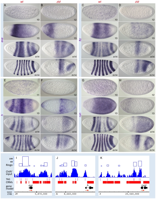
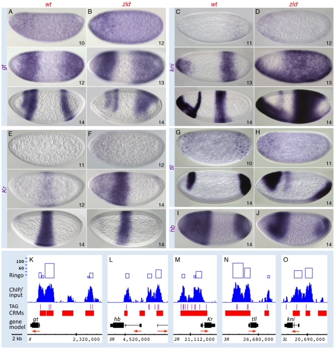
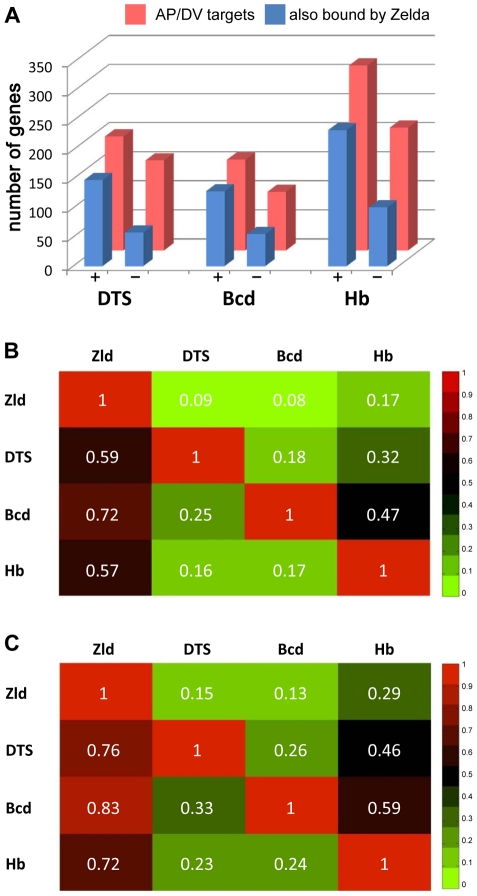
In past years, much attention has focused on the gene networks that regulate early developmental processes, but less attention has been paid to how multiple networks and processes are temporally coordinated. Recently the discovery of the transcriptional activator Zelda (Zld), which binds to CAGGTAG and related sequences present in the enhancers of many early-activated genes in Drosophila, hinted at a mechanism for how batteries of genes could be simultaneously activated. Here we use genome-wide binding and expression assays to identify Zld target genes in the early embryo with the goal of unraveling the gene circuitry regulated by Zld. We found that Zld binds to genes involved in early developmental processes such as cellularization, sex determination, neurogenesis, and pattern formation. In the absence of Zld, many target genes failed to be activated, while others, particularly the patterning genes, exhibited delayed transcriptional activation, some of which also showed weak and/or sporadic expression. These effects disrupted the normal sequence of patterning-gene interactions and resulted in highly altered spatial expression patterns, demonstrating the significance of a timing mechanism in early development. In addition, we observed prevalent overlap between Zld-bound regions and genomic "hotspot" regions, which are bound by many developmental transcription factors, especially the patterning factors. This, along with the finding that the most over-represented motif in hotspots, CAGGTA, is the Zld binding site, implicates Zld in promoting hotspot formation. We propose that Zld promotes timely and robust transcriptional activation of early-gene networks so that developmental events are coordinated and cell fates are established properly in the cellular blastoderm embryo.


Conflict of interest statement

The authors have declared that no competing interests exist.

Figures

Figure 1
Figure 1. Zld protein expression in early embryos.
(A) Time line of the first three hours of Drosophila development representing nc 1 through 14 . Cycle length gradually increases after nc 10 from ten min to one hr at nc 14. Zygotic gene activation begins at 1–2 hrs followed by a larger wave of gene expression at 2–3 hrs. (B) Western analysis of Zld protein in 0–1 hr, 1–2 hr, and 2–3 hr embryos. 40 µg of protein was loaded in each lane and the blot was incubated with anti-Zld antibodies. A protein close to the predicted size of Zld (∼180 kD) increased in concentration after 1 hr of development. (C–F) Zld antibodies detected nuclear protein in wild-type (C–E) but not zld embryos (F, nc 14). Note that Zld protein can be detected as early as nc 2 (C, arrows), and appears to accumulate to higher levels in blastoderm embryos (D, nc 10; E, nc 14).
Figure 2
Figure 2. Conserved TAGteam motifs are differentially enriched in Zld-bound regions.
(A) Enrichment indices (Y-axis) of TAGteam sequences were calculated in 100 bp non-overlapping windows across Zld-bound peaks (5 kb to either side of the center of the peak, marked as 0 on the X-axis). Background was estimated by averaging the enrichment indices of 20 randomly selected heptamers (orange line). (B) PWM derived from the enriched TAGteam sites (see Materials and Methods). (C) PWM derived from the enriched TAT sites (see Materials and Methods). (D) Gel shift assay of labeled oligonucleotides containing different TAGteam sites (lanes 1–16), a mutation of the TAGGTAG site (TGAATAG, lanes 17–18), and the TATCGAT site found in the genome-wide enrichment test (lanes 19–20) without or with recombinant Zld protein (alternating lanes). The eight TAGteam sites bind Zld with varying affinities, while the mutant and TATCGAT site do not bind. (E) Hotspots (3163 with at least eight factors bound) were divided into windows containing 100 hotspots each, and ranked from high to low according to hotspot scores (X-axis, 1 = highest ranking score). The average Zld ChIP/input ratios were calculated for each window (Y-axis). Blue line represents Zld ChIP/input ratios in ranked hotspots; green line represents background (see Materials and Methods).
Figure 3
Figure 3. Zld binds to defined enhancer regions of sc/sisB, zen, and sna.
RNA expression profiles from the 1–2 hr (blue) and 2–3 hr (green) datasets, Zld ChIP/input ratios (binding peaks), and hotspots were viewed on the Integrated Genome Browser (IGB) . All expression peaks are on the same scale (maximum value 15K). Hotspots are shown as orange rectangles; height reflects the hotspot score, which depends on the number of factors bound and the strength of their binding . Zld binding peaks are shown in blue with significance scores from the Ringo algorithm (see Materials and Methods) shown above as blue rectangles. Below the Zld peaks are: TAGteam sites (purple lines; limited to CAGGTAG, TAGGTAG, CAGGTAA), cis-regulatory modules (CRMs from REDfly denoted as filled red boxes , from other sources, denoted as open red boxes), gene models using genome version BDGP R5/dm3 (black rectangles; red arrows denote direction of transcription). Zld binding was observed at the known enhancers of sc/sisB and zen that contain in vivo relevant TAGteam sites, and both the primary and shadow (open red box) enhancers of sna . Peaks were also observed just upstream of the TSS of zen and sna, and to a region downstream of sc/sisB. RNA expression of sc/sisB and zen is mostly absent in zld, while that of sna is less affected. bcd transcripts, which are maternally loaded, are overall similar in wild-type and zld embryos.
Figure 4
Figure 4. Zld potentiates Dl morphogentic activity.
Wild-type (wt; A, C, E, G, I, K) and zld (B, D, F, H, J, L) embryos in nc 11–14 (e, early; m, mid; l, late) were hybridized with RNA probes synthesized against cDNA (A–D) or intronic DNA sequences (see nuclear dots in E–H and I–L insets) for genes indicated on the left. All embryos are oriented anterior to the left and dorsal up, except for the ventral views in E and F (top). (A–H) Nomarksi images. Target-gene transcripts are detectable at nc 11–12 in wild-type embryos, but are delayed by 1–2 nc in zld. By nc 14, twi appears normal (B, bottom), while rho, brk and sog are all restricted to a narrow lateral domain (arrows in D, F, H). (I–L) Confocal FISH images of nc 14 embryos. DAPI stained nuclei expressing rho or sog are shown in yellow. Note the sporadic expression of rho and sog with irregular boundaries in zld. (M) Box plot representing the fraction of active nuclei within the expression domains in the wt vs zld embryos (n = 10–15). Expression domains were variable among zld embryos (data not shown), and showed a significant decrease in the percentage of nuclei (on average 30%) with nascent transcripts (p = 1.02E-11 for rho and p = 1.82E-11 for sog). The expansion of brk dorsally in late nc 14 (D, bottom) is likely due to the absence of dpp in zld. (N–P) IGB views of the genomic regions indicated. About 30 kb of the sog region is not shown (hash marks). Zld-bound regions coincide with known enhancers, including the shadow enhancer of sog (open red box) . See brk primary and shadow enhancers in Figure S1F.
Figure 5
Figure 5. Pair-rule patterns are altered in zld embryos.
Wild-type (wt; A, C, E, G) and zld (B, D, F, H) embryos were hybridized with RNA probes as indicated. Perinuclear transcripts are detectable as early as nc 10; refinement into the seven-stripe patterns occurs in nc 14 (A, C, E, G). In zld, activation is delayed 1–2 cycles (B, D, F, H, top), and refinement is disrupted such that a few aberrant domains develop (B, D, F, H, bottom). (I–K) IGB views of the genomic regions indicated. The ftz region in the Antp complex is shown in Figure S7. Note extensive Zld binding over stripe enhancers and basal promoters. The eve stripe 2 enhancer is located in one of the highest-ranking Zld-bound regions (top 1%, see Figure S3C).
Figure 6
Figure 6. Zld regulates timing within the gap gene network.
Wild-type (wt; A, C, E, G, I) and zld (B, D, F, H, J) embryos were hybridized as indicated. Wild-type activation of gt (A) and tll (G) was detectable earlier than that of kni (C) and Kr (E). In zld embryos, all gap genes were delayed 1–2 nc (B, D, F, H, data not shown for hb), and cross-regulatory interactions were subsequently affected. Abnormal ectopic activation was observed for kni (D) and Kr (F). All gap domains were expanded and/or shifted (B, D, F, H, J), and the gt (B) and Kr (F) domains overlap, which is not seen in wild-type (compare with A and E). (K–O) IGB views of the genomic regions indicated. Multiple Zld-bound regions surround each gap gene (O). Note that the genes activated earlier have higher binding scores (K, N).
Figure 7
Figure 7. Targets of key patterning factors are more likely to be expressed if bound by Zld.
(A) Bar graph showing the number of bound target genes of DV and AP , transcription factors (red) that are also bound by Zld (blue) divided into two groups: targets that are expressed (+) or not expressed (−) in blastoderm embryos (from polII ChIP-chip data of [14]). The analysis was restricted to genes within 2 kb of bound regions. (B–C) Heat maps showing the fraction of target genes that two factors have in common, including all target genes (B) or blastoderm-expressed targets (C). The number in each box represents the fraction of genes bound by a factor denoted in the row that are also bound by the factor denoted in the column. For example, 59% of all DTS targets are also bound by Zld (B). This number increases to 76% for those DTS targets that are expressed at 2–4 hrs (C).

References

    1. Lecuit T, Wieschaus E. Polarized insertion of new membrane from a cytoplasmic reservoir during cleavage of the Drosophila embryo. J Cell Biol. 2000;150:849–860. - PMC - PubMed
    1. ten Bosch JR, Benavides JA, Cline TW. The TAGteam DNA motif controls the timing of Drosophila pre-blastoderm transcription. Development. 2006;133:1967–1977. - PubMed
    1. Liang HL, Nien CY, Liu HY, Metzstein MM, Kirov N, et al. The zinc-finger protein Zelda is a key activator of the early zygotic genome in Drosophila. Nature. 2008;456:400–403. - PMC - PubMed
    1. De Renzis SD, Elemento O, Tavazoie S, Wieschaus EF. Unmasking activation of the zygotic genome using chromosomal deletions in the Drosophila embryo. PLoS Biol. 2007;5:e117. doi: 10.1371/journal.pbio.0050117. - DOI - PMC - PubMed
    1. Roy S, Ernst J, Kharchenko PV, Kheradpour P, Negre N, et al. Identification of functional elements and regulatory circuits by Drosophila modENCODE. Science. 2010;330:1787–1797. - PMC - PubMed

Publication types

MeSH terms