Case Reports
. 2010 Jul;5(2):19-30.

Intracranial Rosai Dorfman Disease: report of three cases and literature review

Affiliations
Case Reports

Intracranial Rosai Dorfman Disease: report of three cases and literature review

Nigel Peter Symss et al. Asian J Neurosurg. 2010 Jul.

Abstract

Background: Rosai-Dorfman Disease (RDD) is a rare idiopathic non-neoplastic histioproliferative disease characterized clinically by massive painless cervical lymphadenopathy, fever and weight loss. Extranodal involvement has also been recognized. Central nervous system (CNS) manifestations are extremely rare and patients with intracranial involvement usually present with clinical and radiological findings suggestive of a meningioma.

Case description: We report our experience in the management of three patients with RDD. Two patients had dural based lesions, radiologically in favour of a meningioma, and one patient had a parenchymal lesion suggestive of a tuberculous granuloma. Treatment consisted of total excision in one case, and subtotal excision followed by conventional radiotherapy in two cases. The diagnosis was confirmed by histopathology and immunochemistry which is essential for a definite diagnosis of RDD.

Keywords: Histoproliferative disease; Lymphnode; Meningioma; Rosai-Dorfman Disease; Tuberculous granuloma.


Figures

Figure 1a
Figure 1a
MRI scan of the brain plain and contrast showing lesion which is isointense in T1 WI, hypointense in T2 WI and uniformly enhancing with contrast. Lesion seen infratentorially extending on both sides along the tentorium, upto the cavernous sinuses, with a larger component on the right side. There is pressure on the right side of the pons, with effacement of the cisterns, and deformity of the fourth ventricle.
Figure 1b
Figure 1b
Post operative CT scan of the brain plain and contrast study showing progression of the lesion bilaterally.
Figure 1c
Figure 1c
Follow up CT scan of the brain at eight years showing progression in size of the lesion with extending into the cavernous sinuses upto the orbit.
Figure 2a
Figure 2a
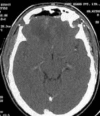
MRI of the brain showed a bifrontal dural based lesion with involvement of the falx. There is perilesional oedema and mass effect.
Figure 2b
Figure 2b
MRI of the brain with gadolinium showing intense contrast enhancement of the bifrontal dural based lesion.
Figure 2c
Figure 2c
Post operative CT scan of the brain showing total excision of the lesion.
Figure 3a
Figure 3a
MRI scan of the brain showing an ill-defined contrast enhancing lesion in the suprasellar region.
Figure 3b
Figure 3b
Repeat MRI scan of the brain plain study and with gadolinium showing multiple ring enhancing lesions in the sellar, suprasellar, retrosellar and interpeduncular regions, with intra-axial lesion in the left medial temporal region, with surrounding vasogenic oedema, and mass effect
Figure 3c
Figure 3c
Repeat MRI scan of the brain plain and gadolinium showing confluent ring enhancing lesions in the suprasellar, retrosellar and interpeduncular regions, with intraaxial ring enhancing lesions in the left temporal, subfrontal regions, upper pons and right side of lower midbrain. There is extensive surrounding vasogenic oedema, with mass effect on the frontal and temporal horns, with dilatation of the lateral ventricles and third ventricle.
Figure 3d
Figure 3d
Magnetic resonance spectroscopy showing elevated lipid and N-Acetyl Aspartate peaks suggestive of a granulomatous lesion.
Figure 3e
Figure 3e
Post operative CT scan of the brain showing multiple ill defined hyperdense, contrast enhancing residual lesion in the sellar, suprasellar, retrosellar, and interpeduncular regions, left basifrontal and left temporal lobe with perilesional oedema and mass effect on the lateral ventricles, with post operative changes.
Figure 3f
Figure 3f
Follow up CT scan of the brain plain and contrast study showing further resolution of the lesion.

References

    1. Andriko JA, Morrison A, Colegial CH, et al. Rosai-Dorfman disease isolated to the central nervous system: A report of 11 cases. Mod Pathol. 2001;14:172–8. - PubMed
    1. Cheng-Hong Toh, Yao-Liang Chen, Ho-Fai Wong, et al. Rosai—Dorfman disease with dural sinus invasion Report of two cases. J Neurosurg. 2005;102:550–554. - PubMed
    1. Deodhare SS, Ang LC, Bilbao JM. Isolated intracranial involvement in Rosai- Dorfman disease.A report of two cases and review of the literature. Arch Pathol Lab Med. 1998;122:161–5. - PubMed
    1. Destombes P. Adenitis avec surcharge lipidique, de l’ enfant ou de l’adulte jeune, odservees aux Antilles et au Mali. Bull Soc Pathol Exot. 1965;6:1169–75. - PubMed
    1. Fabrice Bing, Jean-Paul Brion, Sylvie Grand, et al. Tumor arising in the periventricular region. Neuropathology. 2009;29:101–103. - PubMed

Publication types

LinkOut - more resources