

Robust reproducible resting state networks in the awake rodent brain
- PMID: 22028788
- PMCID: PMC3196498
- DOI: 10.1371/journal.pone.0025701
Robust reproducible resting state networks in the awake rodent brain
Abstract
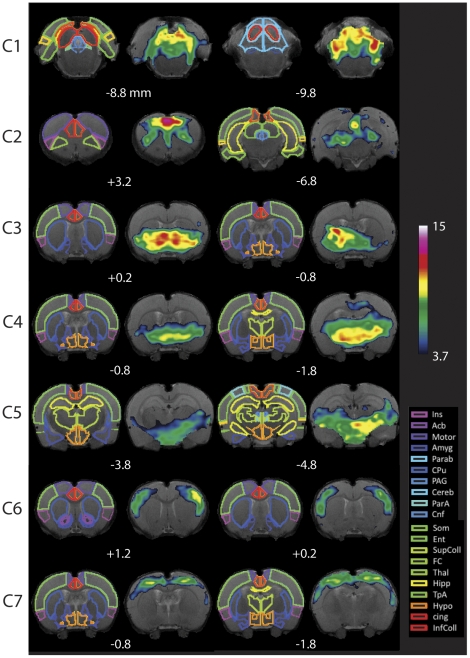
Resting state networks (RSNs) have been studied extensively with functional MRI in humans in health and disease to reflect brain function in the un-stimulated state as well as reveal how the brain is altered with disease. Rodent models of disease have been used comprehensively to understand the biology of the disease as well as in the development of new therapies. RSN reported studies in rodents, however, are few, and most studies are performed with anesthetized rodents that might alter networks and differ from their non-anesthetized state. Acquiring RSN data in the awake rodent avoids the issues of anesthesia effects on brain function. Using high field fMRI we determined RSNs in awake rats using an independent component analysis (ICA) approach, however, ICA analysis can produce a large number of components, some with biological relevance (networks). We further have applied a novel method to determine networks that are robust and reproducible among all the components found with ICA. This analysis indicates that 7 networks are robust and reproducible in the rat and their putative role is discussed.
Conflict of interest statement
Figures
References
-
- Damoiseaux JS, Beckmann CF, Arigita EJ, Barkhof F, Scheltens P, et al. Reduced resting-state brain activity in the “default network” in normal aging. Cereb Cortex. 2008;18:1856–1864. - PubMed
-
- Auer DP. Spontaneous low-frequency blood oxygenation level-dependent fluctuations and functional connectivity analysis of the ‘resting’ brain. Magn Reson Imaging. 2008;26:1055–1064. - PubMed
-
- van den Heuvel MP, Hulshoff Pol HE. Exploring the brain network: a review on resting-state fMRI functional connectivity. Eur Neuropsychopharmacol. 2010;20:519–534. - PubMed
-
- Biswal B, Yetkin FZ, Haughton VM, Hyde JS. Functional connectivity in the motor cortex of resting human brain using echo-planar MRI. Magn Reson Med. 1995;34:537–541. - PubMed
Publication types
MeSH terms
Grants and funding
LinkOut - more resources
Full Text Sources
Miscellaneous
