. 2011;6(10):e26137.
doi: 10.1371/journal.pone.0026137. Epub 2011 Oct 10.

Salivary PYY: a putative bypass to satiety

Affiliations

Salivary PYY: a putative bypass to satiety

Andres Acosta et al. PLoS One. 2011.

Abstract

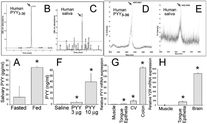
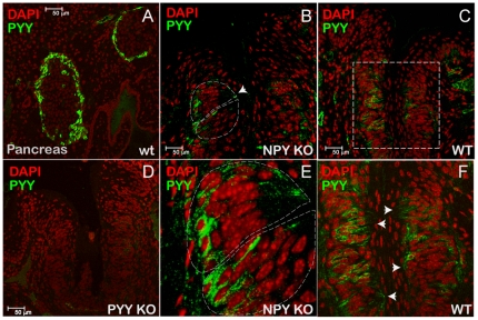
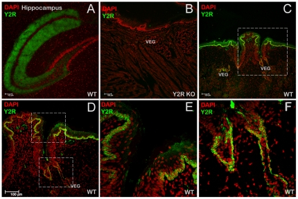
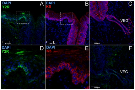
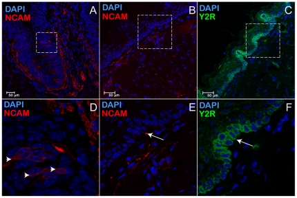
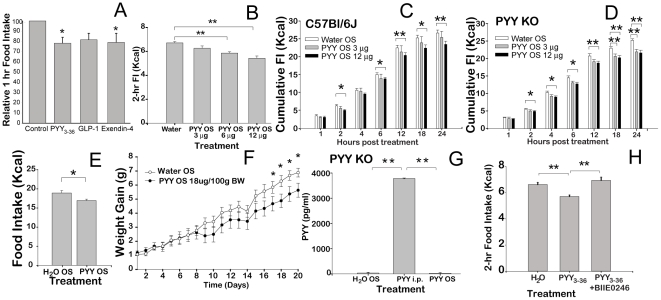
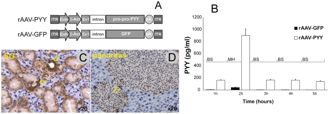
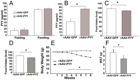
Peptide YY(3-36) is a satiation hormone released postprandially into the bloodstream from L-endocrine cells in the gut epithelia. In the current report, we demonstrate PYY(3-36) is also present in murine as well as in human saliva. In mice, salivary PYY(3-36) derives from plasma and is also synthesized in the taste cells in taste buds of the tongue. Moreover, the cognate receptor Y2R is abundantly expressed in the basal layer of the progenitor cells of the tongue epithelia and von Ebner's gland. The acute augmentation of salivary PYY(3-36) induced stronger satiation as demonstrated in feeding behavioral studies. The effect is mediated through the activation of the specific Y2 receptor expressed in the lingual epithelial cells. In a long-term study involving diet-induced obese (DIO) mice, a sustained increase in PYY(3-36) was achieved using viral vector-mediated gene delivery targeting salivary glands. The chronic increase in salivary PYY(3-36) resulted in a significant long-term reduction in food intake (FI) and body weight (BW). Thus this study provides evidence for new functions of the previously characterized gut peptide PYY(3-36) suggesting a potential simple and efficient alternative therapeutic approach for the treatment of obesity.


Conflict of interest statement

Competing Interests: The authors have declared that no competing interests exist.

Figures

Figure 1
Figure 1. Characterization of PYY3-36 origins in human and of murine saliva.
(A) Concentration of PYY3-36 in human saliva during fasting and 30 minutes after of finishing a standardized meal (n = 5), *P<0.05. (B) Separation of synthetic PYY3-36 by RP-HPLC. (C) Separation of human saliva by RP-HPLC. (D) Validation of synthetic PYY3-36 by MALDI-TOF (E) Validation of PYY3-36 from human saliva by MALDI-TOF. Arrows indicate PYY3-36 peaks. (F) PYY3-36 in saliva from male C57Bl/6J mice (n = 10 per group) injected with saline or synthetic murine PYY3-36 i.p., at 10 min post injection. (G) RT-PCR assay measuring relative PYY mRNA expression in the muscle as a (-) control, tongue epithelium, and circumvallate papillae (CV) with colon as a (+) control in male C57Bl/6 mice (n = 10 per group). (H) RT-PCR assay measuring relative Y2R mRNA expression in the tongue epithelium, muscle as a (-) control, and brain as a (+) control in male C57Bl/6 mice (n = 8 per group). *P<0.05.
Figure 2
Figure 2. PYY is synthesized in taste cells.
(A) Immunolocalization of PYY-positive cells in a-cells in the murine pancreas, a (+) control. (B) Immunolocalization of PYY in CV of a NPY KO mouse, a control for PYY antibodies cross-reactivity. (C) Immunolocalization of PYY in CV of a C57Bl/6J mouse (WT). (D) Immunolocalization of PYY in CV of a PYY KO mouse, a (-) control. (E) close-up of (B). (F) close-up of (C). Arrowheads point at the apical part of a taste bud.
Figure 3
Figure 3. Y2 receptor is synthesized in the epithelial cells of the tongue.
(A) Immunolocalization of Y2R-positive cells in the hippocampus of C57Bl/6J mouse (WT), a (+) control. (B) Immunolocalization of Y2R in the tongue epithelia of Y2R KO mouse, a (-) control. VEG – von Ebner's gland. (C) Immunolocalization of Y2R-positive cells in the CV area of the tongue of a C57Bl/6J mouse. (D) close-up of (C). (E), and (F) close ups of (D), top and bottom rectangles, respectively.
Figure 4
Figure 4. A subpopulation of epithelial progenitor cells in the tongue epithelia expresses Y2R.
Two sequential mirror sections of the tongue were immunostained for Y2R (A, D, and and F), or Cytokeratin 5 (K5) (B, E, and C). For better viewing, K5 images were reflected horizontally. Areas at the sulcus edge, positive for both Y2R (A) and K5 (B) (dashed rectangles), are shown as close-up images in (D) and (E), respectively. Panels (C) and (F) show Y2R and K5-positive cells in von Ebner's gland connecting to CV's sulcus.
Figure 5
Figure 5. Neuronal filaments innervate circumvallate papillae (CV) as well as the basal layer of cells distant from CV.
Immunostaining for NCAM in CV (A) shows subpopulation of taste cells expressing K5 (marked by arrowheads in panel D, a close-up from panel A), as well as a dense mesh of filaments at the basolateral surfaces of the taste buds. Rectangles in (B) and (C) designate the same areas in two sequential mirror sections stained for NCAM (red), or Y2R (green). The protrusions in the tongue epithelia surface (B, C, E, and F) are filiform papillae transversely sectioned. Even distant from CV, the Y2R-positive epithelial layer is morphologically close to neuro-filament layer below (E). Some Y2R cells and NCAM filaments appear to be juxtaposed (arrows in E and F).
Figure 6
Figure 6. Oral PYY3-36 augmentation therapy.
(A) Effect of PYY3-36 (3 mg/100 g BW), GLP-1(3 mg/100 g BW), or Exendin-4 (10 mg/100 g BW) oral spray (n = 10 each group) on 1 hour FI after 24 hours fasting compared to control oral spray (n = 10). Experiments for each peptide were conducted separately and the results were integrated in one graph as fractions of average amount of FI consumed by the control group. PYY3-36-, and Exendin-4-treated mice consumed significantly less food. (B) Dose-response effect of PYY3-36 on 2 hr FI vs. control (n = 8 each group). (C) Effect of PYY3-36 oral spray on FI in C57Bl/6J mice measured at 1, 2, 6, 12, 18, and 24 hr post treatment (n = 8/group). (D) Effect of PYY3-36 oral spray on FI in PYY-/- mice measured at 1, 2, 6, 12, 18, and 24 hr post treatment (n = 8/group). (E) Average 24-hr FI in DIO mice treated with daily PYY3-36 OS (18 mg/100 g). (F) Effect of daily PYY3-36 OS (18 mg/100 g) treatment on BW change in DIO C57Bl/6J mice (n = 9 per each group). (G) Concentration of PYY3-36 in plasma of PYY KO mice 10 min after PYY3-36 (18 mg/100 g BW), or control oral spray vs. PYY3-36 injected i.p. (6 mg/100 g BW) (n = 10 per group). (H) Effect of Y2R specific antagonist BIIE0246 on anorexigenic action of PYY3-36 (n = 8 per each group) measured as 2 hr FI after 24 hr fast.*P<0.05, **P<0.01.
Figure 7
Figure 7. Validation of recombinant Adeno-associated virus encoding pre-pro-PYY (rAAV-PYY).
(A) Diagram of rAAV-PYY and rAAV-GFP cassettes: ITR - inverted terminal repeats of rAAV serotype 2; CBA - Cytomegalovirus intermediate early enhancer sequence/chicken b-acting promoter; murine Pre-pro-Peptide YY cDNA, GFP - green fluorescence protein cDNA. (B) PYY secretion study in NCI-H716 cells. The experiment was performed on 3 different occasions with 10 wells per group. Media was assayed at the times indicated. BS – basal secretion; MH – secretion stimulated with 2% meat hydrolysate. (C) Cellular distribution of rAAV-PYY in salivary glands. Submandibular glands infused with rAAV-PYY removed 22 weeks after administration and evaluated for PYY expression by immunohistochemistry. The brown color indicates PYY-positive cells. Arrows indicate PYY-positive striated ducts. (D) Positive control – PYY-stained cells in murine pancreas.
Figure 8
Figure 8. Effect of PYY gene transfer to the salivary glands in C57Bl/6J mice.
(A) Concentration of PYY3-36 in plasma during fasting and after feeding. (B) Concentration of PYY3-36 in saliva. (C) Effect of PYY3-36 salivary gland gene delivery on weekly FI in mice fed HF diet. (D) Effect of PYY3-36 salivary gland gene delivery on weekly FI in DIO mice. (E) BW change in DIO C57Bl/6J treated with rAAV-GFP (control group) and rAAV-PYY over 8 weeks post injection. (F) Weight of visceral white adipose tissue at 8 weeks post injection in mice treated with rAAV (all groups were 10 animals/group) *P<0.05.

References

    1. Vallejo G, Mead PM, Gaynor DH, Devlin JT, Robbins DC. Characterization of immunoreactive insulin in human saliva: evidence against production in situ. Diabetologia. 1984;27:437–440. - PubMed
    1. Groschl M, Rauh M, Wagner R, Neuhuber W, Metzler M, et al. Identification of leptin in human saliva. J Clin Endocrinol Metab. 2001;86:5234–5239. - PubMed
    1. Toda M, Tsukinoki R, Morimoto K. Measurement of salivary adiponectin levels. Acta Diabetol. 2007;44:20–22. - PubMed
    1. Shin YK, Martin B, Golden E, Dotson CD, Maudsley S, et al. Modulation of taste sensitivity by GLP-1 signaling. J Neurochem. 2008;106:455–463. - PMC - PubMed
    1. Herness MS. Vasoactive intestinal peptide-like immunoreactivity in rodent taste cells. Neuroscience. 1989;33:411–419. - PubMed

Publication types

MeSH terms