. 2011;6(10):e26288.
doi: 10.1371/journal.pone.0026288. Epub 2011 Oct 18.

Attenuated inflammatory response in aged mice brains following stroke

Affiliations

Attenuated inflammatory response in aged mice brains following stroke

Matthias W Sieber et al. PLoS One. 2011.

Abstract

Background: Increased age is a major risk factor for stroke incidence, post-ischemic mortality, and severe and long-term disability. Stroke outcome is considerably influenced by post-ischemic mechanisms. We hypothesized that the inflammatory response following an ischemic injury is altered in aged organisms.

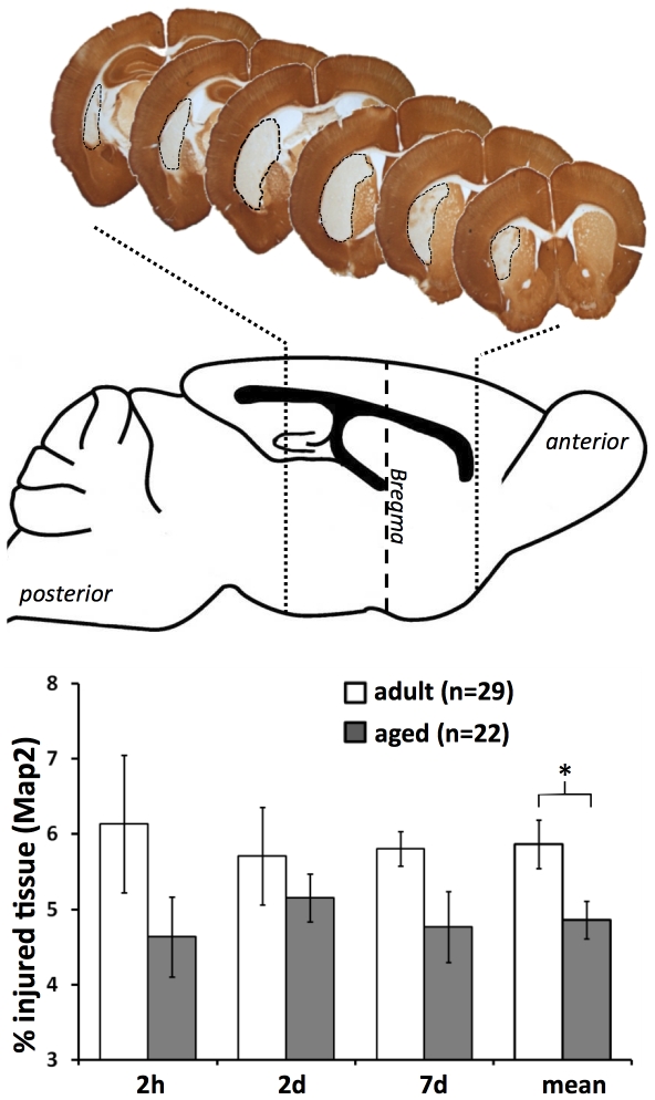
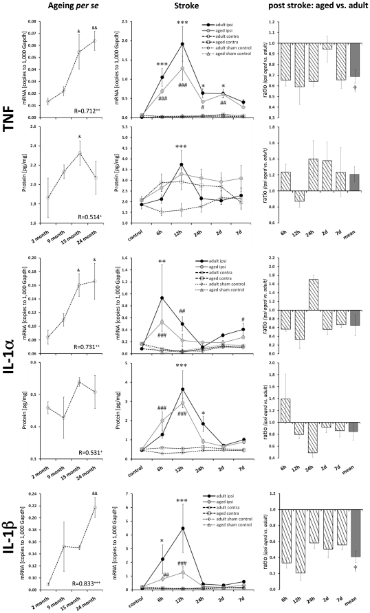
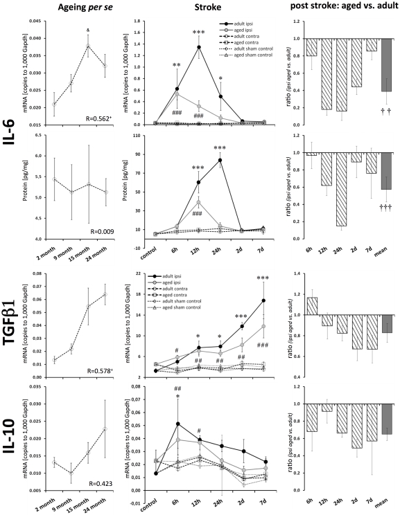
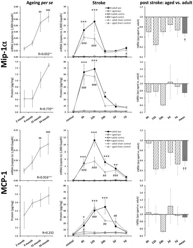
Methods and results: To that end, we analyzed the expression pattern of pro-inflammatory cytokines (TNF, IL-1α, IL-1β, IL-6), anti-inflammatory cytokines (IL-10, TGFβ1), and chemokines (Mip-1α, MCP-1, RANTES) of adult (2 months) and aged (24 months) mice brains at different reperfusion times (6 h, 12 h, 24 h, 2 d, 7 d) following transient occlusion of the middle cerebral artery. The infarct size was assessed to monitor possible consequences of an altered inflammatory response in aged mice. Our data revealed an increased neuro-inflammation with age. Above all, we found profound age-related alterations in the reaction to stroke. The response of pro-inflammatory cytokines (TNF, and IL-1β) and the level of chemokines (Mip-1α, and MCP-1) were strongly diminished in the aged post-ischemic brain tissue. IL-6 showed the strongest age-dependent decrease in its post-ischemic expression profile. Anti-inflammatory cytokines (TGFβ1, and IL-10) revealed no significant age dependency after ischemia. Aged mice brains tend to develop smaller infarcts.

Conclusion: The attenuated inflammatory response to stroke in aged animals may contribute to their smaller infarcts. The results presented here highlight the importance of using aged animals to investigate age-associated diseases like stroke, and should be considered as a major prerequisite in the development of age-adjusted therapeutic interventions.


Conflict of interest statement

Competing Interests: The authors have declared that no competing interests exist.

Figures

Figure 1
Figure 1. Infarct volumes at different ages.
The mean infarct volume (±SEM) was significantly smaller in aged mice brains (Map2 immunohistochemistry, ANOVA with post-hoc Tukey test, * p≤0.05).
Figure 2
Figure 2. Age- and stroke-dependent expression of TNF, IL-1α, and IL-1β.
TNF, IL-1α, and IL-1β displayed significantly elevated expression levels with age. All these pro-inflammatory cytokines showed a distinct response following stroke, which was attenuated in aged brains. Expression values are shown as mean ± SEM and ratios as geometric mean ± SEM. Significant age-related differences (versus 2-month-old native mice) are indicated by & p≤0.05, && p≤0.01, and &&& p≤0.001 (ANOVA and post-hoc Tukey test). Correlation of the cytokine expression level with age is indicated by + p≤0.05, ++ p≤0.01, and +++ p≤0.001 (R, Pearson correlation). Significant post-stroke differences (ipsi versus contra) are indicated by adult: * p≤0.05, ** p≤0.01, and *** p≤0.001; and aged: # p≤0.05, ## p≤0.01, and ### p≤0.001 (paired two-way ANOVA and post-hoc Tukey test). Significant age-dependent differences after stroke (ipsi aged versus ipsi adult) are displayed by † p≤0.05, †† p≤0.01, and ††† p≤0.001 (two-way ANOVA).
Figure 3
Figure 3. Age- and stroke-dependent expression of IL-6, TGFβ1 and IL-10.
IL-6 transcript expression significantly correlated with age; however, this correlation was not reflected in the expression of its protein. The post-ischemic IL-6 response was attenuated in aged brains (mRNA and protein). TGFβ1 mRNA significantly correlated with age. IL-10 mRNA tended to be elevated in aged brains. TGFβ1 transcript expression in the ischemic tissue increased continuously with reperfusion time at both ages. The up-regulation of TGFβ1 and IL-10 following ischemia tended to be attenuated in aged brains. Expression values are shown as mean ± SEM and ratios as geometric mean ± SEM. For details of the statistical analysis, see Figure 2's legend or the Materials and Methods section.
Figure 4
Figure 4. Age- and stroke-dependent expression of Mip-1α and MCP-1.
Mip-1α and MCP-1 displayed significantly elevated expression levels with age. Mip-1α and MCP-1 transcript and protein expression increased significantly following stroke. This up-regulation was attenuated in aged brains. Expression values are shown as mean ± SEM and ratios as geometric mean ± SEM. For details of the statistical analysis, see Figure 2's legend or the Materials and Methods section.
Figure 5
Figure 5. Age- and stroke-dependent expression of RANTES.
The expression of RANTES significantly correlated with age. After ischemia, the level of RANTES mRNA continuously increased up to 7 d in adult brains, whereas no up-regulation was observed in aged brains. Protein levels of RANTES were up-regulated in adult and aged brains. Expression values are shown as mean ± SEM and ratios as geometric mean ± SEM. For details of the statistical analysis, see Figure 2's legend or the Materials and Methods section.
Figure 6
Figure 6. Schematic diagram summarizing the post-ischemic expression profiles of all the tested inflammatory mediators.
The relative expression profiles (%) of all the tested inflammatory mediators in adult (continuous line, colored area) and aged brains (dotted line, light colored area) are shown (mRNA, left panel; protein, right panel). The post-ischemic response of TNF, IL-1α, IL-1β, IL-6, Mip-1α, MCP-1, and TGFβ1 was considerably attenuated with age, with IL-6 exhibiting the strongest age-dependent effect.

References

    1. Dilger RN, Johnson RW. Aging, microglial cell priming, and the discordant central inflammatory response to signals from the peripheral immune system. J Leukoc Biol. 2008;84:932–939. - PMC - PubMed
    1. Truelsen T, Piechowski-Jozwiak B, Bonita R, Mathers C, Bogousslavsky J, et al. Stroke incidence and prevalence in Europe: a review of available data. Eur J Neurol. 2006;13:581–598. - PubMed
    1. Donnan GA, Fisher M, Macleod M, Davis SM. Stroke. Lancet. 2008;371:1612–1623. - PubMed
    1. Saposnik G, Black SE, Hakim A, Fang J, Tu JV, et al. Age disparities in stroke quality of care and delivery of health services. Stroke. 2009;40:3328–3335. - PubMed
    1. O'Collins VE, Macleod MR, Donnan GA, Horky LL, van der Worp BH, et al. 1,026 experimental treatments in acute stroke. Ann Neurol. 2006;59:467–477. - PubMed

Publication types