. 2011;6(10):e26298.
doi: 10.1371/journal.pone.0026298. Epub 2011 Oct 18.

Ectopic PDX-1 expression directly reprograms human keratinocytes along pancreatic insulin-producing cells fate

Affiliations

Ectopic PDX-1 expression directly reprograms human keratinocytes along pancreatic insulin-producing cells fate

Michal Mauda-Havakuk et al. PLoS One. 2011.

Abstract

Background: Cellular differentiation and lineage commitment have previously been considered irreversible processes. However, recent studies have indicated that differentiated adult cells can be reprogrammed to pluripotency and, in some cases, directly into alternate committed lineages. However, although pluripotent cells can be induced in numerous somatic cell sources, it was thought that inducing alternate committed lineages is primarily only possible in cells of developmentally related tissues. Here, we challenge this view and analyze whether direct adult cell reprogramming to alternate committed lineages can cross the boundaries of distinct developmental germ layers.

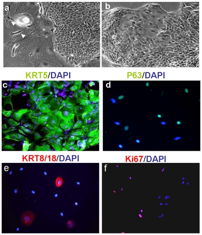
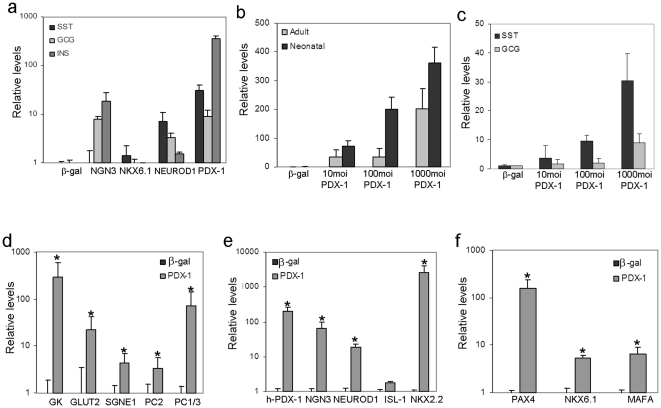
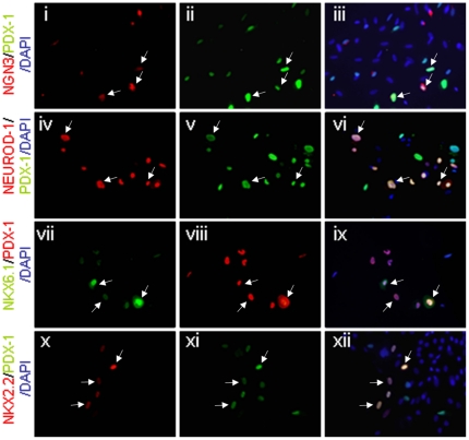
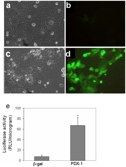
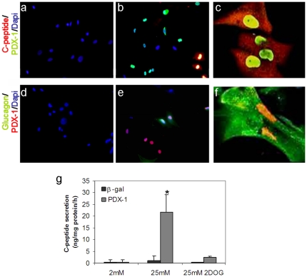
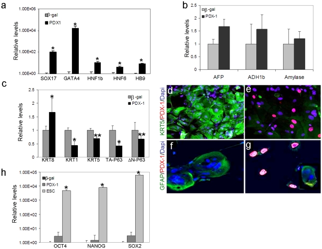
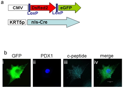
Methodology/principal findings: We ectopically expressed non-integrating pancreatic differentiation factors in ectoderm-derived human keratinocytes to determine whether these factors could directly induce endoderm-derived pancreatic lineage and β-cell-like function. We found that PDX-1 and to a lesser extent other pancreatic transcription factors, could rapidly and specifically activate pancreatic lineage and β-cell-like functional characteristics in ectoderm-derived human keratinocytes. Human keratinocytes transdifferentiated along the β cell lineage produced processed and secreted insulin in response to elevated glucose concentrations. Using irreversible lineage tracing for KRT-5 promoter activity, we present supporting evidence that insulin-positive cells induced by ectopic PDX-1 expression are generated in ectoderm derived keratinocytes.

Conclusions/significance: These findings constitute the first demonstration of human ectoderm cells to endoderm derived pancreatic cells transdifferentiation. The study represents a proof of concept which suggests that transcription factors induced reprogramming is wider and more general developmental process than initially considered. These results expanded the arsenal of adult cells that can be used as a cell source for generating functional endocrine pancreatic cells. Directly reprogramming somatic cells into alternate desired tissues has important implications in developing patient-specific, regenerative medicine approaches.


Conflict of interest statement

Competing Interests: The authors have declared that no competing interests exist.

Figures

Figure 1
Figure 1. Characterization of primary culture of human keratinocytes in-vitro.
Human keratinocytes proliferated efficiently for up to 5–7 passages in culture. (a, b) Representative phase contrast images show the morphology of keratinocytes at passages (a) 0, and (b) 3. Arrowheads (a) indicate cells of the 3T3 fibroblast feeder layer in passage 0; original magnification ×10. (c–f) Keratinocytes (passage 3) were stained for keratinocyte markers: (c) KRT5, (d) P63, (e) KRT8/18, and (f) for the proliferation marker, Ki67. Nuclei were stained with DAPI (blue); original magnifications, ×20.
Figure 2
Figure 2. Ectopic PDX-1 expression promoted pancreatic differentiation in human keratinocytes.
(a) Human keratinocytes isolated from neonatal foreskin were analyzed by quantitative RT-PCR to determine insulin (INS), glucagon (GCG), and somatostatin (SST) gene transcription levels induced by adenoviral infections with transcription factor genes PDX-1, NGN3, NKX6.1, or NEUROD1. The control virus carried only the β-gal gene sequence. All vectors were infected at 1000 moi and cells were analyzed 7 days post infection. (b) Human keratinocytes isolated from neonatal foreskin or adult skin grafts, (9–70 year-old donors) were infected with increasing concentrations of PDX-1 (10–1000 moi) or control (β-gal) adenovirus constructs to determine INS gene transcription. (c) Adult keratinocytes were infected with increasing concentrations of PDX-1 (10–1000 moi) or control (β-gal) adenovirus constructs to determine GCG and SST gene transcription levels. (d, e, f) Keratinocytes from neonatal foreskin were infected with PDX-1 or control (β-gal) adenovirus constructs (both at 1000 moi) to determine transcription levels of genes encoding (d) pancreatic-specific proteins, (e) pancreatic specific transcription factors, and (f) β-cell specific transcription factors. The results are normalized to β-actin gene transcription within the same cDNA samples. The mean ±standard deviations are expressed as the signal strength relative to cells treated with the control virus. All results represent n≥8 samples tested in three independent experiments *P<0.01.
Figure 3
Figure 3. Nuclear localization of PDX-1 induced pancreatic transcription factor expression in human keratinocytes.
Human keratinocytes isolated from neonatal foreskin were analyzed 7 days subsequent to Ad-PDX-1 infection (ectopic PDX-1). Double Immunofluorescence analyses showed co-localization of (i, ii, iii) NGN3 (red) and ectopic PDX-1 (green); (iv, v, vi) NEUROD1 (red) and ectopic PDX-1 (green); (vii, vii, ix) NKX6.1 (green) and ectopic PDX-1 (red); and (x, xi, xii) NKX2.2 (red) and ectopic PDX-1 (green). In panels iii, vi, ix, and xii, the images were superimposed to visualize co-localization (merged colors). Nuclei were stained with DAPI (blue). Original magnification ×20; arrows indicate double-stained cells.
Figure 4
Figure 4. Ectopic PDX-1 expression activated the insulin promoter in human keratinocytes in-vitro.
Human keratinocytes isolated from neonatal foreskin were co-infected with either the control virus Ad-β-gal or Ad-PDX-1 (both at 1000 moi) and a reporter gene of either (a–d) Ad-RIP-GFP (1000 moi,) or (e) Ad-RIP-Luciferase (100 moi,), for 7 days. (a, c) Representative phase contrast micrographs show cell morphology. (b, d) Green fluorescent protein imaging of the same fields (a&b; c&d) presented in the phase contrast. (b) control cells did not express GFP; and (d) PDX-1 stimulated GFP expression in numerous cells. (e) Luciferase activity of the infected keratinocytes. The mean ±standard deviation is expressed in relative luciferase units; data represents n≥8 samples in three independent experiments.
Figure 5
Figure 5. Ectopic PDX-1 expression induced insulin and glucagon expression and promoted glucose-regulated C-peptide secretion.
Double immunofluorescence of human keratinocytes isolated from neonatal foreskin treated with (a, d) control Ad-β-gal or (b–c, e–f) Ad-PDX-1 adenovirus for 7 days. Immunofluorescence analyses showed co-localized expression of (a–c) PDX-1(green) and insulin (red) and (d–f) PDX-1(red) and glucagon (green). The nuclei were stained with DAPI (blue). (a–b, d–e). Original magnification ×20 (c, f); original magnification (confocal microscope) ×90. (g) Human keratinocytes were infected with Ad-β-gal or Ad-PDX-1, both at 1000 moi, and analyzed for secretion of processed insulin into the culture medium upon exposure to 2 and 25 mM glucose or 25 mM 2DOG. C-peptide in the medium was measured with a specific RIA kit (Linco human c-peptide no crossreactivity with pro-insulin). Data represents n≥7 samples in four independent experiments.
Figure 6
Figure 6. Ectopic PDX-1 activated early endoderm but not hepatic or pancreatic acinar markers, while repressing the expression of keratinocyte-specific markers.
Human keratinocytes isolated from neonatal foreskin were infected with Ad-β-gal or Ad-PDX-1 (both at 1000 moi) for 7 days. Quantitative real time RT-PCR analyses indicated the relative transcription levels of (a) early endoderm-specific markers, (b) hepatic and pancreatic acinar markers, (c) keratinocyte markers or (h) embryonic stem cell markers. The results were normalized to the level of β-actin gene transcription within the same cDNA sample. The mean ±standard deviations of Ad-PDX-1 treated cells are expressed relative to the mean levels observed in Ad-β-gal treated cells. n≥6 from 3 different cultures. *P<0.01, **P<0.05 (h) cDNA extracted from human embryonic cells was used as positive control. (d–g) Cells treated with either (d, f) Ad-β-gal or (e, g) Ad-PDX-1 were fixed and co-stained for (d, e) KRT5 (green) and PDX-1 (red) or for (f, g) GFAP (green) and PDX-1 (red). The nuclei were stained with DAPI; magnification: d–g ×30.
Figure 7
Figure 7. C-peptide positive cells arose from keratinocytes (KRT5 positive cells).
Lineage tracing analyses were performed with dual Cre-Lox lentiviral infections. (a) Schematic representation of the Lox (upper) and Cre (lower) lentiviral vectors. Only cells that expressed KRT5 could activate Cre expression; then, Cre would excise the DsRed2 gene between the Lox sites and remove a stop codon preceding the eGFP gene, which is subsequently controlled by the CMV promoter. (b) Ten days after the dual lentiviral infection, cells were treated with Ad-PDX-1 for 7 days. Fixed cells were stained with three immunofluorescent antibodies and examined with a confocal microscope to detect keratinocytes that processed insulin. (i, ii, iii) GFP (green) identifies the cell as a keratinocyte, PDX-1(blue) shows successful ectopic protein expression, and c-peptide (turquoise) shows processed insulin production. (iv) Superimposed images show that all three proteins are expressed within the same cell. Original magnification ×90.

References

    1. Raff M. Adult stem cell plasticity: fact or artifact? Annu Rev Cell Dev Biol. 2003;19:1–22. - PubMed
    1. Jopling C, Boue S, Izpisua Belmonte JC. Dedifferentiation, transdifferentiation and reprogramming: three routes to regeneration. Nature. 2011;12:79–89. - PubMed
    1. Theise ND. New principles of cell plasticity. C R Biol. 2002;325:1039–1043. - PubMed
    1. Theise ND. Stem cell plasticity: recapping the decade, mapping the future. Exp. 2010;38:529–539. - PubMed
    1. Hanna J, Markoulaki S, Schorderet P, Carey BW, Beard C, et al. Direct reprogramming of terminally differentiated mature B lymphocytes to pluripotency. Cell. 2008;133:250–264. - PMC - PubMed

Publication types

MeSH terms