. 2011;6(10):e26397.
doi: 10.1371/journal.pone.0026397. Epub 2011 Oct 20.

Human engineered heart tissue as a versatile tool in basic research and preclinical toxicology

Affiliations

Human engineered heart tissue as a versatile tool in basic research and preclinical toxicology

Sebastian Schaaf et al. PLoS One. 2011.

Abstract

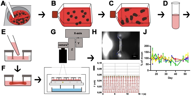
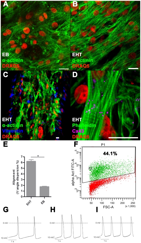
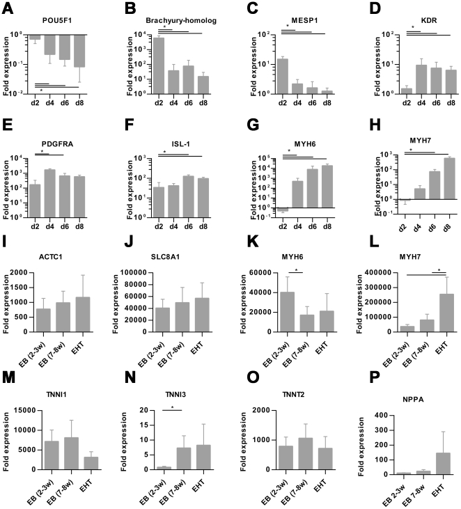
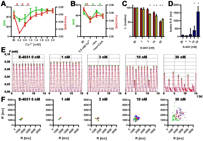
Human embryonic stem cell (hESC) progenies hold great promise as surrogates for human primary cells, particularly if the latter are not available as in the case of cardiomyocytes. However, high content experimental platforms are lacking that allow the function of hESC-derived cardiomyocytes to be studied under relatively physiological and standardized conditions. Here we describe a simple and robust protocol for the generation of fibrin-based human engineered heart tissue (hEHT) in a 24-well format using an unselected population of differentiated human embryonic stem cells containing 30-40% α-actinin-positive cardiac myocytes. Human EHTs started to show coherent contractions 5-10 days after casting, reached regular (mean 0.5 Hz) and strong (mean 100 µN) contractions for up to 8 weeks. They displayed a dense network of longitudinally oriented, interconnected and cross-striated cardiomyocytes. Spontaneous hEHT contractions were analyzed by automated video-optical recording and showed chronotropic responses to calcium and the β-adrenergic agonist isoprenaline. The proarrhythmic compounds E-4031, quinidine, procainamide, cisapride, and sertindole exerted robust, concentration-dependent and reversible decreases in relaxation velocity and irregular beating at concentrations that recapitulate findings in hERG channel assays. In conclusion this study establishes hEHT as a simple in vitro model for heart research.


Conflict of interest statement

Competing Interests: The authors have declared that no competing interests exist.

Figures

Figure 1
Figure 1. Schematic illustration of cardiac differentiation, EHT generation and analysis.
A: Undifferentiated hESC colonies are detached after collagenase treatment with 5 ml pipette; B: EBs are formed in ultra-low attachment flasks (for details see Figure S1). C: Cardiomyocytes are differentiated and onset of beating in EBs is monitored; D: EBs are enzymatically dissociated into single cells and a mastermix (cells, fibrinogen, thrombin) is prepared; E: Mastermix is pipetted into EHT casting molds; F: Within two weeks coherent beating EHTs develop under cell culture conditions between silicone posts; G: EHTs in 24 well plates are analyzed by automated video-optical recording, therefore a camera is placed above the incubator and directed to each well by x-y-z-coordinate motor system; H: Movies are recorded, automated figure recognition modus identifies top and bottom end of each EHT (blue squares); I: Based on post deflection, post geometry and elastic propensity force development is calculated and recorded over time. Contraction peaks are automatically recognized (green squares) and parameters of contraction are calculated and summarized in a report; J: Force (red), frequency (green), contraction velocity (blue), relaxation velocity (yellow) of 4 EHTs between day 15 and day 55 of development, lines show means ± SEM.
Figure 2
Figure 2. Immunofluorescence, FACS, electrophysiology.
A: Immunofluorescence staining of 2–3 week old EB, B–D: Immunofluorescence staining of EHT, scale bar 20 µm; E: Quantification of alignment: In EHT format cardiomyocytes are significantly more aligned than in EB-format *P<0.05 (Student's t-test), bars show mean ± SD. F: Representative FACS analysis of dissociated EBs (2–3 weeks) before EHT generation, P1: All gated cells, P2: α-actinin positive cells. Representative recordings of APs: G: EBs (2–3 weeks), H: EBs (7–8 weeks), I: EHT (5 weeks): 7–8 week old EBs as well as EHT derived cardiomyocytes have longer APSs compared to young EB derived cardiomyocytes. Furthermore the maximal diastolic potential is particularly low in EHT derived cardiomyocytes.
Figure 3
Figure 3. Transcript analysis.
A–H: Quantitative RT-PCR analysis of stemness marker (POU5F1, POU class 5 homeobox 1), mesodermal marker (brachyury homolog, MESP1, mesoderm posterior 1 homolog, KDR, vascular endothelial growth factor receptor 2, PDGFRA, platelet derived growth factor receptor alpha) and cardiac marker (ISL1, Islet-1, MYH6, α-myosin heavy chain, MYH7, ß-myosin heavy chain,) in EB-based cardiac differentiation on day 2, 4, 6, and 8, normalized to undifferentiated hESCs. For these experiments Stage 1 duration (mesodermal induction) was set to 24 hours, 4–7 biological replicas. I–L: Quantitative RT-PCR analysis of alpha actin (ACTC1), sodium/calcium exchanger (SLC8A1), MYH6 and MYH7 in EBs (2–3 weeks old), EBs (7–8 weeks old) and EHTs (5 weeks old) normalized to undifferentiated hESC, 4–6 biological replica. Bars show mean ± SD, *P<0.05 (Student's t-test), bars show means ± SD.
Figure 4
Figure 4. Functional analysis of hEHTs.
A and B: Calcium concentration-response curve: Development of force of contraction (red, mN) and frequency (green, beat per minute, BPM) and baseline (Bl) and increasing Ca2+ concentration (A), Response to isoprenaline and carbachol (B). C: Analysis of force and relaxation velocity in the presence of E-4031. Red: force, yellow: relaxation velocity, depicted as percent of baseline values. *P<0.05 (Student's t-test), 4 biological replica, bars show means ± SD. D: Scatter of beat-to-beat variability in the presence of E-4031, *P<0.05 (Mann-Whitney U test), 4 biological replica, bars show median ± interquartile range. E: Original recordings (15 sec each) of spontaneous EHT contractions under increasing concentrations of E-4031 (0–30 nM). F: Statistical evaluation of beat-to-beat variability of the experiments depicted under E. Ordinates indicates the distance from a given twitch to the following, the abscissa the distance to the previous twitch. Biological replicas are discriminated by color code.

References

    1. Kehat I, Kenyagin-Karsenti D, Snir M, Segev H, Amit M, et al. Human embryonic stem cells can differentiate into myocytes with structural and functional properties of cardiomyocytes. The Journal of Clinical Investigation. 2001;108:407–14. - PMC - PubMed
    1. Yang L, Soonpaa MH, Adler ED, Roepke TK, Kattman SJ, et al. Human cardiovascular progenitor cells develop from a KDR+ embryonic-stem-cell-derived population. Nature. 2008;453:524–8. - PubMed
    1. Mummery C, Ward-van Oostwaard D, Doevendans P, Spijker R, Brink S van den, et al. Differentiation of human embryonic stem cells to cardiomyocytes: role of coculture with visceral endoderm-like cells. Circulation. 2003;107:2733–40. - PubMed
    1. Xu XQ, Graichen R, Soo SY, Balakrishnan T, Rahmat SNB, et al. Chemically defined medium supporting cardiomyocyte differentiation of human embryonic stem cells. Differentiation. 2008;76:958–70. - PubMed
    1. Laflamme M a, Chen KY, Naumova AV, Muskheli V, Fugate J, et al. Cardiomyocytes derived from human embryonic stem cells in pro-survival factors enhance function of infarcted rat hearts. Nature Biotechnology. 2007;25:1015–24. - PubMed

MeSH terms