. 2011;6(10):e26504.
doi: 10.1371/journal.pone.0026504. Epub 2011 Oct 18.

Osteoblast-specific transcription factor Osterix increases vitamin D receptor gene expression in osteoblasts

Affiliations

Osteoblast-specific transcription factor Osterix increases vitamin D receptor gene expression in osteoblasts

Chi Zhang et al. PLoS One. 2011.

Abstract

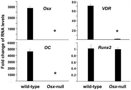
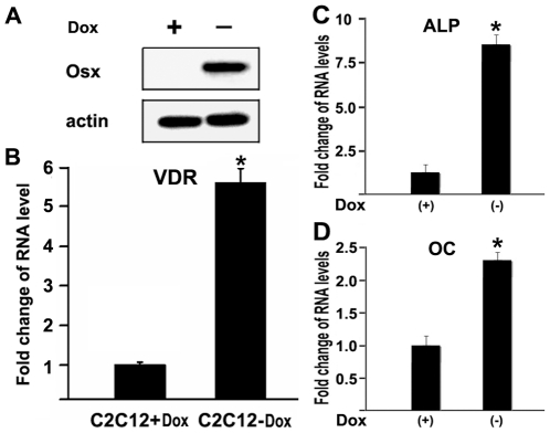
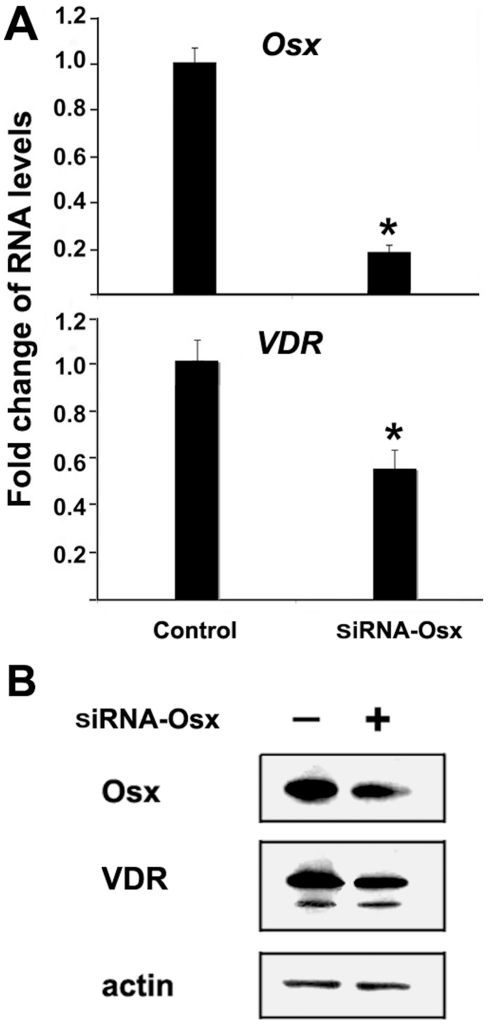
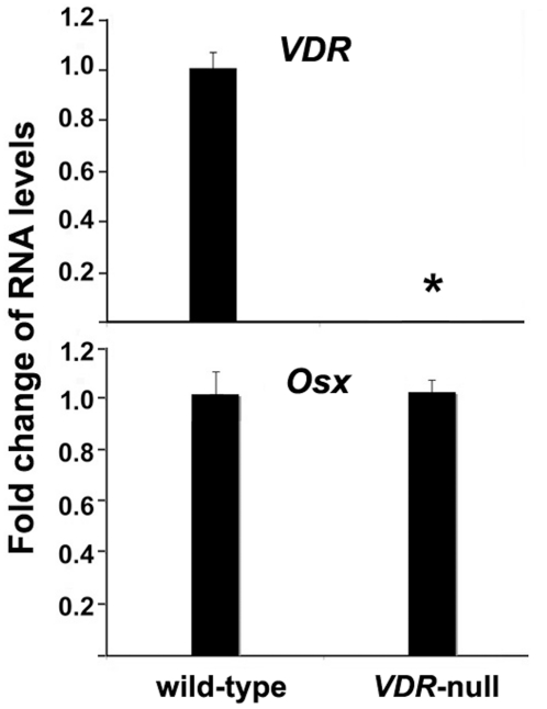
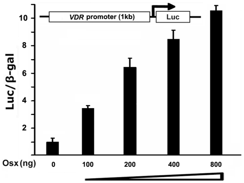
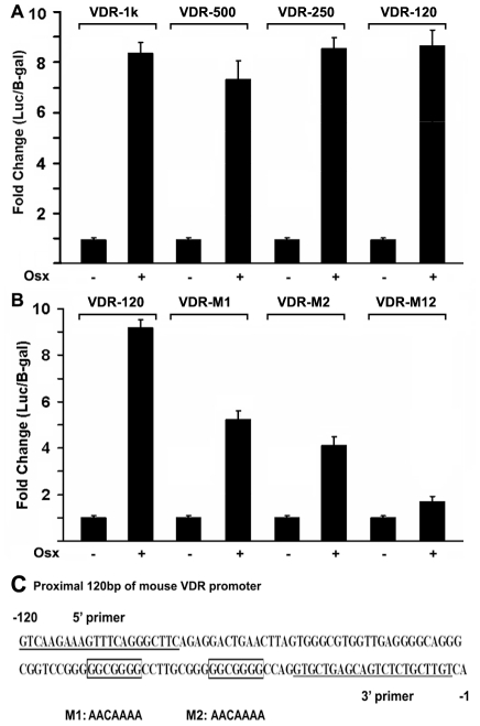
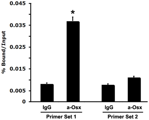
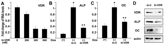
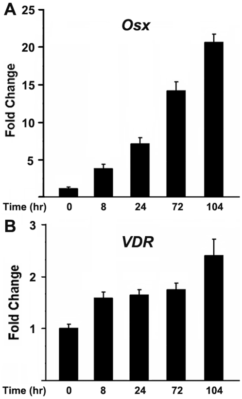
Osterix (Osx) is an osteoblast-specific transcription factor required for osteoblast differentiation from mesenchymal stem cells. In Osx knock-out mice, no bone formation occurs. The vitamin D receptor (VDR) is a member of the nuclear hormone receptor superfamily that regulates target gene transcription to ensure appropriate control of calcium homeostasis and bone development. Here, we provide several lines of evidence that show that the VDR gene is a target for transcriptional regulation by Osx in osteoblasts. For example, calvaria obtained from Osx-null embryos displayed dramatic reductions in VDR expression compared to wild-type calvaria. Stable overexpression of Osx stimulated VDR expression in C2C12 mesenchymal cells. Inhibition of Osx expression by siRNA led to downregulation of VDR. In contrast, Osx levels remained unchanged in osteoblasts in VDR-null mice. Mechanistic approaches using transient transfection assays showed that Osx directly activated a 1 kb fragment of the VDR promoter in a dose-dependent manner. To define the region of the VDR promoter that was responsive to Osx, a series of VDR promoter deletion mutants were examined and the minimal Osx-responsive region was refined to the proximal 120 bp of the VDR promoter. Additional point mutants were used to identify two GC-rich regions that were responsible for VDR promoter activation by Osx. Chromatin immunoprecipitation assays demonstrated that endogenous Osx was associated with the native VDR promoter in primary osteoblasts in vivo. Cumulatively, these data strongly support a direct regulatory role for Osx in VDR gene expression. They further provide new insight into potential mechanisms and pathways that Osx controls in osteoblasts and during the process of osteoblastic cell differentiation.


Conflict of interest statement

Competing Interests: The authors have declared that no competing interests exist.

Figures

Figure 1
Figure 1. Osx ablation reduces VDR gene expression in vivo.
Calvaria RNAs were isolated from E18.5 Osx wild-type and Osx-null embryos. RNA expression levels for Osx, osteocalcin (OC), Runx2 and VDR were analyzed by real-time RT-PCR. The level of each RNA from Osx-null calvaria was normalized to a value of 1. *: A star indicates statistical significance compared to Osx wild type group.
Figure 2
Figure 2. Overexpression of Osx activates VDR gene expression in C2C12 mesenchymal cells.
(A) Western immunoblot analysis of the Dox-regulated Osx-expressing C2C12 cells. Osx expression is turned on in the absence of Dox. Beta-actin was used as a loading control. (B) VDR mRNA levels in a stable, Tet-off C2C12 mesenchymal cell line. RNA was obtained from cultures treated with or without Doxycycline. Osx expression is induced in the absence of Doxycycline in this line. VDR mRNA levels were quantitated by real-time RT-PCR. The VDR RNA level obtained from the cells cultured with Dox was normalized to a value of 1. Values are presented as the mean ±S.D. (C) RNA expression level for the osteoblastic marker gene alkaline phosphatase (ALP). (D) RNA expression level for osteoblastic gene osteocalcin (OC). Conditions are identical to those described in panel B. A paired t-test was performed comparing Dox (−) and Dox (+) groups. *: A star indicates statistical significance compared to Dox (+) group.
Figure 3
Figure 3. SiRNA-directed knockdown of Osx impairs VDR gene expression in MC3T3 osteoblasts.
(A) RNA expression levels as determined by quantitative real-time RT-PCR. MC3T3 osteoblasts were transfected with siRNA targeting mouse Osx. RNA was isolated 24 h post-transfection and quantitated by real-time RT-PCR. The RNA level from the control siRNA group was normalized to a value of 1. Values were presented as the mean ±S.D. A paired t-test was performed comparing si-control group and si-Osx group. (B) Western analysis of the Osx knockdown. Protein was isolated by acetone precipitation of whole cell lysates and then analyzed by western blot using rabbit anti-VDR or anti-Osx polyclonal antibodies. Beta-actin was used as a loading control.
Figure 4
Figure 4. VDR ablation has no effect on Osx gene expression in vivo.
Calvarial RNAs were isolated from E18.5 wild-type or VDR-null embryos. RNA levels for VDR and Osx were analyzed by real-time RT-PCR. The level of each RNA from wild type calvaria was normalized to a value of 1 shown in the mean±S.D. A paired t-test was performed comparing wild-type and VDR-null group.
Figure 5
Figure 5. Osx activates the VDR promoter in a dose-dependent manner.
HEK293 cells were transfected with a 1 kb VDR promoter-luciferase reporter gene without or with increasing amounts of an Osx-expression plasmid as indicated. Luciferase activity was normalized by β-galactosidase activity. Values are presented as the mean ±S.D.
Figure 6
Figure 6. Identification of the Osx binding site in the promoter of VDR gene.
(A) Deletion analysis of the VDR promoter-reporter construct. VDR-1 kb, VDR-500 bp, VDR-250 bp and VDR-120 bp promoter-reporter plasmids (300 ng each) were cotransfected with 400 ng of the Osx expression plasmid in HEK293 cells. Twenty-four hours post-transfection, cell extracts were prepared and analyzed for luciferase activity and normalized to β-galactosidase activity. (B) Two GC-rich elements in VDR-120 are responsible for VDR promoter reporter activation by Osx. The promoter mutants VDR-M1, VDR-M2 and VDR-M12 were transfected into HEK293 cells and analyzed as described in panel A. Luciferase activity was normalized by β-galactosidase activity. (C) A diagram of the proximal 120 bp region of the mouse VDR promoter. A 5′ primer and 3′ primer were used to subclone the VDR-120 bp promoter reporter plasmid. M1 refers to point mutations of VDR-M1, and M2 refers to point mutations of VDR-M2. VDR-M12 in (B) contains both M1 and M2.
Figure 7
Figure 7. Endogenous Osx in primary osteoblasts is associated with the native VDR promoter in vivo.
Chromatin Immunoprecipitation (ChIP) assays were conducted using primary calvarial osteoblasts isolated from new born wild-type mice. Anti-Osx antibody (a-Osx) was used for ChIP analysis, and IgG was used as a negative control. The precipitated chromatin was analyzed by quantitative real-time PCR. As described in the Methods, primer Set 1 corresponds to a segment covering two GC-rich elements within 120 bp the VDR promoter. As a negative control, Primer Set 2 covers a distal 3 kb region of the VDR promoter, which does not contain GC-rich sequences. A paired t-test was performed comparing IgG and a-Osx group.
Figure 8
Figure 8. Osx-regulated expression of VDR is important in Osx-induced osteoblast differentiation.
(A) SiRNA directed against VDR knocks down VDR expression. C2C12 mesenchymal cells stably expressing Osx were transfected with VDR siRNA at different concentrations. RNA was isolated and specific RNA levels were measured by real-time RT-PCR. (B,C) Cells were transfected with 300 nM of siRNA targeting VDR, and Osx expression was induced by removing Dox. Osteoblast marker gene expression was examined including (B) ALP and (C)osteocalcin (OC). The RNA level from control siRNA group (Si-C) was normalized to a value of 1. Values were presented as the mean ±S.D. *: A star indicates statistical significance compared to Dox (+) group (p<0.05). **: Two stars indicate statistical significance compared to si-control (Si-C) (p<0.05). (D) Western analysis of the VDR knockdown. C2C12 stable cells without Dox were transfected with 300 nM of siRNA targeting VDR. Protein was isolated from whole cell lysates of control siRNA group (si-C) and VDR siRNA group (si-VDR) and then analyzed by western blot using rabbit anti-VDR, anti-ALP or anti-OC polyclonal antibodies. Beta-actin was used as a loading control.
Figure 9
Figure 9. Both Osx and VDR are upregulated during osteoblast differentiation.
MC3T3 osteoblast differentiation experiments were performed in which osteogenic factors were added into the medium, including BMP2, ascorbic acid and β–glycerophosphate. MC3T3 osteoblastic cells were harvested after 0 hr, 8 hr, 24 hr, 72 hr and 104 hr after incubating with differentiation medium. RNA was isolated from cell lysates. RNA levels for VDR and Osx were analyzed by real-time RT-PCR. The level of RNA from 0 hr was normalized to a value of 1. Values were presented as the mean ±S.D.

References

    1. Zhang C. Transcriptional regulation of bone formation by the osteoblast-specific transcription factor Osx. J Orthop Surg Res. 2010;5:37. - PMC - PubMed
    1. St-Jacques B, Hammerschmidt M, McMahon AP. Indian hedgehog signaling regulates proliferation and differentiation of chondrocytes and is essential for bone formation. Genes Dev. 1999;13:2072–2086. - PMC - PubMed
    1. Komori T, Yagi H, Nomura S, Yamaguchi A, Sasaki K, et al. Targeted disruption of Cbfa1 results in a complete lack of bone formation owing to maturational arrest of osteoblasts. Cell. 1997;89:755–764. - PubMed
    1. Nakashima K, Zhou X, Kunkel G, Zhang Z, Deng JM, et al. The novel zinc finger-containing transcription factor osterix is required for osteoblast differentiation and bone formation. Cell. 2002;108:17–29. - PubMed
    1. Rodda SJ, McMahon AP. Distinct roles for Hedgehog and canonical Wnt signaling in specification, differentiation and maintenance of osteoblast progenitors. Development. 2006;133:3231–3244. - PubMed

Publication types