. 2011;6(10):e26514.
doi: 10.1371/journal.pone.0026514. Epub 2011 Oct 20.

The transcription factors Snail and Slug activate the transforming growth factor-beta signaling pathway in breast cancer

Affiliations

The transcription factors Snail and Slug activate the transforming growth factor-beta signaling pathway in breast cancer

Archana Dhasarathy et al. PLoS One. 2011.

Abstract

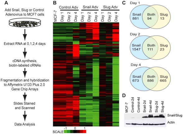
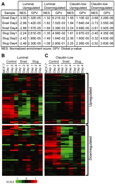
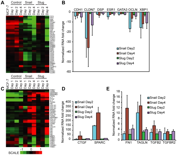
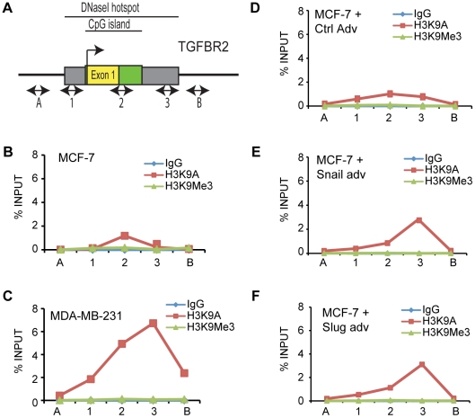
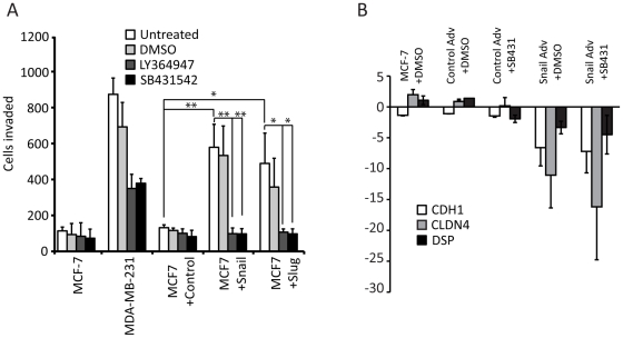
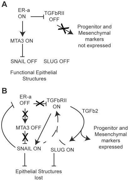
The transcriptional repressors Snail and Slug are situated at the core of several signaling pathways proposed to mediate epithelial to mesenchymal transition or EMT, which has been implicated in tumor metastasis. EMT involves an alteration from an organized, epithelial cell structure to a mesenchymal, invasive and migratory phenotype. In order to obtain a global view of the impact of Snail and Slug expression, we performed a microarray experiment using the MCF-7 breast cancer cell line, which does not express detectable levels of Snail or Slug. MCF-7 cells were infected with Snail, Slug or control adenovirus, and RNA samples isolated at various time points were analyzed across all transcripts. Our analyses indicated that Snail and Slug regulate many genes in common, but also have distinct sets of gene targets. Gene set enrichment analyses indicated that Snail and Slug directed the transcriptome of MCF-7 cells from a luminal towards a more complex pattern that includes many features of the claudin-low breast cancer signature. Of particular interest, genes involved in the TGF-beta signaling pathway are upregulated, while genes responsible for a differentiated morphology are downregulated following Snail or Slug expression. Further we noticed increased histone acetylation at the promoter region of the transforming growth factor beta-receptor II (TGFBR2) gene following Snail or Slug expression. Inhibition of the TGF-beta signaling pathway using selective small-molecule inhibitors following Snail or Slug addition resulted in decreased cell migration with no impact on the repression of cell junction molecules by Snail and Slug. We propose that there are two regulatory modules embedded within EMT: one that involves repression of cell junction molecules, and the other involving cell migration via TGF-beta and/or other pathways.


Conflict of interest statement

Competing Interests: The authors have declared that no competing interests exist. Please note that the authors Dhiral Phadke, Deepak Mav and Ruchir R Shah are employed by a commercial company, SRA International Inc., and performed data analysis under contract from NIEHS. This does not alter the authors' adherence to all the PLoS ONE policies on sharing data and materials.

Figures

Figure 1
Figure 1. Snail and Slug regulate common and unique sets of genes in breast cancer cells.
A) Schematic representation of microarray experiment. B) Heat map depicting probes with absolute fold >2 and adjusted p-value<0.01 on Day4 after Snail and Slug expression, shown over time. C) Venn diagrams showing the total number of genes that change following Snail or Slug expression, with absolute fold >2 and adjusted p-value<0.01. D) Immunoblots showing the expression of Snail, Slug or Actin in MCF-7 (untreated) and following 2–4 days of adenovirus expression.
Figure 2
Figure 2. Snail and Slug cause changes in expression that most resemble the claudin-low class of breast tumors.
GSEA analysis (A) comparing the genes in the microarray samples to those that were upregulated (B and C, top panels) or downregulated (B and C, bottom panels) from the claudin-low class (B) and luminal class (C) described in . See text for details. NES = Normalized enrichment score, GPV = Global p-value.
Figure 3
Figure 3. Snail and Slug expression in MCF-7 cells decreases the expression of differentiation markers, and increases the expression of TGF-beta pathway genes.
Heat maps depicting (A) differentiated cell markers (C) TGF-b pathway probes following Snail and Slug expression. (B, D and E) RT-PCR of cDNA with real-time quantitation following normalization to 18 s, MCF-7 Day 0 and control adenovirus. The data represents the average of three independent biological replicates.
Figure 4
Figure 4. TGFBR2 shows distinct histone acetylation patterns in MCF7 and MDA231, and this changes upon addition of Snail and Slug.
Primers for ChIP were designed across the TGFBR2 promoter region (A). Chromatin IP across the TGFBR2 promoter in MDA-MB-231 (C) demonstrated an increased amount of histone H3K9 acetylation compared to MCF-7 cells (B). Increased acetylation is also seen when Snail or Slug are expressed in MCF-7 cells (E and F), relative to the control-adenovirus infected cells (D). The data is representative of three independent biological replicates.
Figure 5
Figure 5. Treatment with a TGF-beta inhibitor reduces the migratory response to Snail and Slug.
(A) Cell migration assay using MDA-MB-231, uninfected MCF-7 cells, and MCF-7 cells infected with control, Snail or Slug adenoviruses (white bars), treated with DMSO (light grey bars) or with 10 µM final of the TGF-beta inhibitors LY364947 (dark grey bars) or SB431542 (black bars) for 2 days. The X-axis represents the total number of cells in 10 fields. The data represents the average of three independent biological replicates (* T-test p-value ≤0.05, **T-test p-value ≤0.01). (B) Addition of TGF-beta inhibitor does not affect repression of cell junction molecules CDH1, DSP and CLDN4. MCF-7 cells were untreated or treated with control or Snail adenovirus, and with DMSO vehicle or SB431542 for 2 days. RNA was isolated and RT-PCR of cDNA with real-time quantitation was performed following normalization to GAPDH, and MCF-7 Day 0. The data represents the average of three independent biological replicates.
Figure 6
Figure 6. A role for Snail and Slug in upregulating TGF-beta during EMT.
In normal breast cells, the TGF-beta pathway is active in keeping Estrogen receptor-alpha cells from proliferating. A) In cancer cells with a luminal phenotype (e.g. MCF-7), the TGF-beta pathway is downregulated, ER-alpha and MTA3 are expressed, and Snail expression is inhibited. This results in a differentiated and epithelial phenotype with well-preserved cell junctions, and the cells are adherent and non-migratory. B) During EMT, expression of Snail (and/or Slug) causes downregulation of ER-alpha and upregulation of the TGF-beta pathway. However, TGF-beta now serves a pro-metastatic role, and early progenitor markers are induced, resulting in a more ‘claudin-low’ morphology that is more mesenchymal and highly migratory.

References

    1. Polyak K. Breast cancer: origins and evolution. J Clin Invest. 2007;117:3155–3163. - PMC - PubMed
    1. Blick T, Hugo H, Widodo E, Waltham M, Pinto C, et al. Epithelial mesenchymal transition traits in human breast cancer cell lines parallel the CD44(hi/)CD24 (lo/-) stem cell phenotype in human breast cancer. J Mammary Gland Biol Neoplasia. 2010;15:235–252. - PubMed
    1. Trimboli AJ, Fukino K, de Bruin A, Wei G, Shen L, et al. Direct evidence for epithelial-mesenchymal transitions in breast cancer. Cancer Res. 2008;68:937–945. - PubMed
    1. Nieto MA. The snail superfamily of zinc-finger transcription factors. Nat Rev Mol Cell Biol. 2002;3:155–166. - PubMed
    1. Cano A, Perez-Moreno MA, Rodrigo I, Locascio A, Blanco MJ, et al. The transcription factor snail controls epithelial-mesenchymal transitions by repressing E-cadherin expression. Nat Cell Biol. 2000;2:76–83. - PubMed

Publication types

MeSH terms