. 2011;6(10):e26658.
doi: 10.1371/journal.pone.0026658. Epub 2011 Oct 19.

Human evolution and osteoporosis-related spinal fractures

Affiliations

Human evolution and osteoporosis-related spinal fractures

Meghan M Cotter et al. PLoS One. 2011.

Abstract

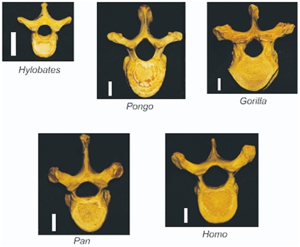
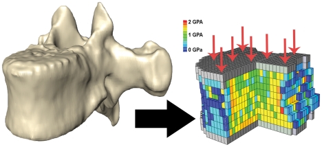
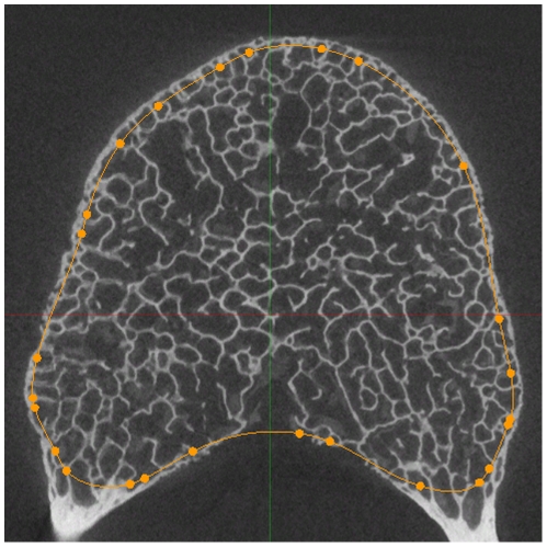
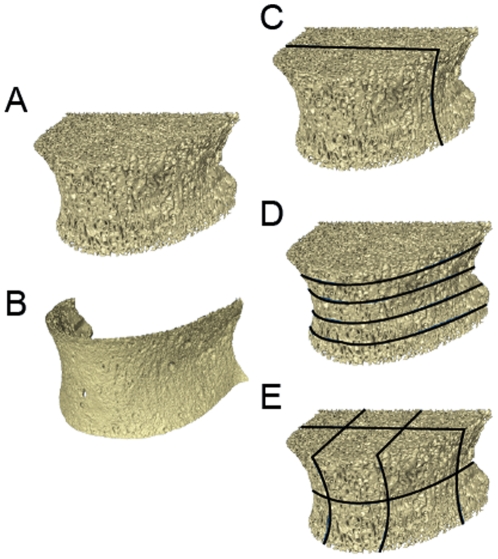
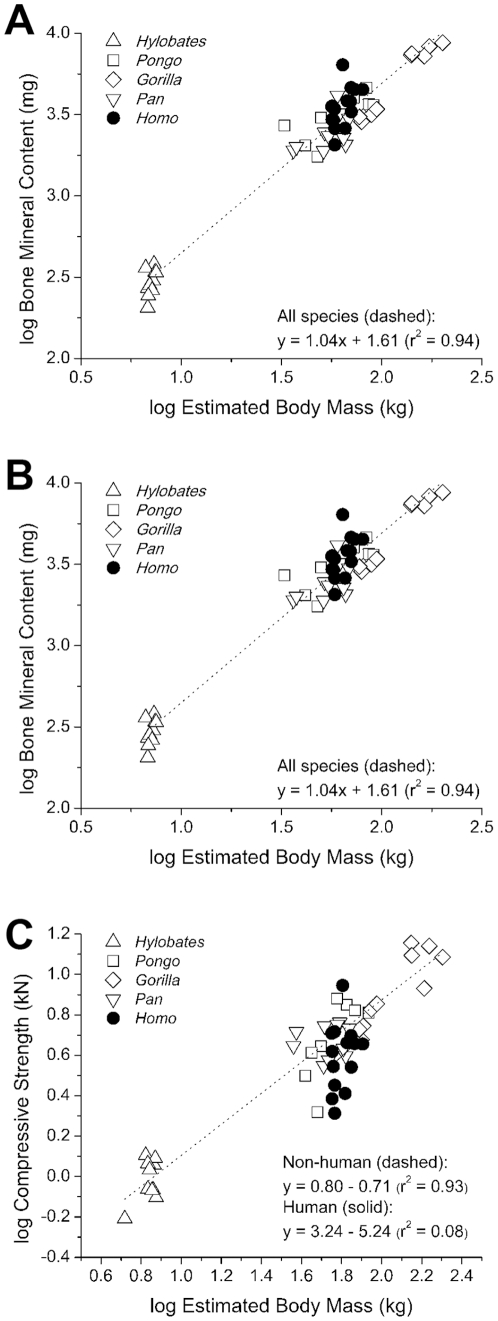
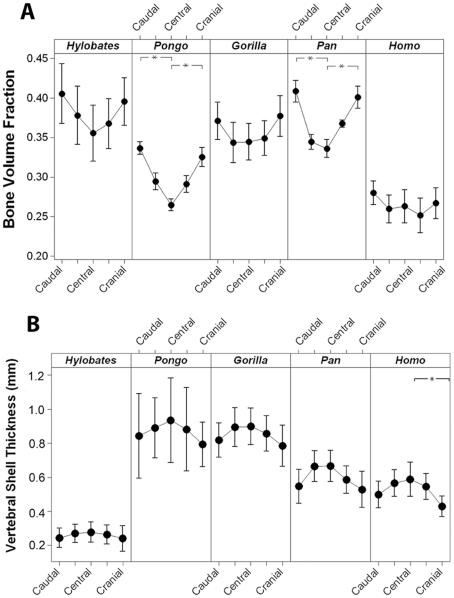
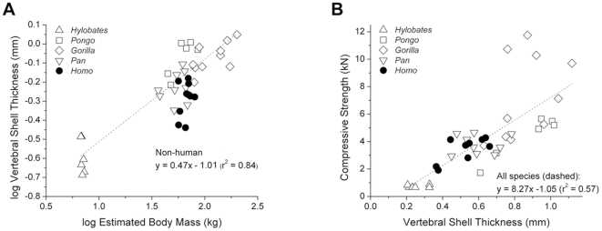
The field of evolutionary medicine examines the possibility that some diseases are the result of trade-offs made in human evolution. Spinal fractures are the most common osteoporosis-related fracture in humans, but are not observed in apes, even in cases of severe osteopenia. In humans, the development of osteoporosis is influenced by peak bone mass and strength in early adulthood as well as age-related bone loss. Here, we examine the structural differences in the vertebral bodies (the portion of the vertebra most commonly involved in osteoporosis-related fractures) between humans and apes before age-related bone loss occurs. Vertebrae from young adult humans and chimpanzees, gorillas, orangutans, and gibbons (T8 vertebrae, n = 8-14 per species, male and female, humans: 20-40 years of age) were examined to determine bone strength (using finite element models), bone morphology (external shape), and trabecular microarchitecture (micro-computed tomography). The vertebrae of young adult humans are not as strong as those from apes after accounting for body mass (p<0.01). Human vertebrae are larger in size (volume, cross-sectional area, height) than in apes with a similar body mass. Young adult human vertebrae have significantly lower trabecular bone volume fraction (0.26±0.04 in humans and 0.37±0.07 in apes, mean ± SD, p<0.01) and thinner vertebral shells than apes (after accounting for body mass, p<0.01). Since human vertebrae are more porous and weaker than those in apes in young adulthood (after accounting for bone mass), even modest amounts of age-related bone loss may lead to vertebral fracture in humans, while in apes, larger amounts of bone loss would be required before a vertebral fracture becomes likely. We present arguments that differences in vertebral bone size and shape associated with reduced bone strength in humans is linked to evolutionary adaptations associated with bipedalism.


Conflict of interest statement

Competing Interests: The authors have declared that no competing interests exist.

Figures

Figure 1
Figure 1. Specimens examined in the study.
The study included thoracic vertebrae from five genera including Hylobates (gibbons), Pongo (orangutan), Gorilla, Pan (chimpanzee) and Homo (humans). The scale bar next to each representative specimen is one centimeter in length.
Figure 2
Figure 2. Biomechanical modeling to determine bone strength.
(A) Three-dimensional images of vertebral bodies (a chimpanzee vertebra shown) were converted into finite element models for biomechanical analysis. (B) Finite element models were loaded in compression (arrows). Differences in the color of the bone elements represent different regional density and elastic modulus.
Figure 3
Figure 3. Delineation of vertebral shell and trabecular bone boundary.
In transverse cross-sectional slices of the vertebrae, the boundary between the vertebral shell and the trabecular bone was traced. Here, an orange line denotes that boundary in a human vertebral body. Characteristics of the bone such as orientation of the bone and relative thickness of the shell and trabecular bone helped determine the placement of the boundary.
Figure 4
Figure 4. Analysis of trabecular microarchitecture.
A micro-computed tomography image of a human vertebral body is shown. Images were divided into (A) trabecular bone and (B) vertebral shell. Variation in trabecular microarchitecture within the vertebral body was examined by considering variation in microarchitecture in (C) dorsal-ventral subregions, (D) transverse subregions and (E) across 12 anatomically determined subregions.
Figure 5
Figure 5. The relationships between body mass, bone mass and bone strength.
(A) A positive correlation between body mass and bone mass (measured as bone mineral content) of the T8 vertebral body was observed that was similar in all species. (B) A positive correlation between bone mass and compressive strength of the T8 vertebral body was observed. Vertebrae from humans displayed reduced strength relative to bone mass (ANCOVA: p<0.01). (C) Although body mass was positively correlated with bone strength across species, vertebrae from humans showed reduced strength as compared to apes with similar body mass (ANCOVA: p = 0.04).
Figure 6
Figure 6. Cranial-Caudal variation in trabecular bone volume fraction and shell thickness.
Interval plots displaying mean (filled circle) and 95% confidence interval (bars) of (A) bone volume fraction and (B) vertebral shell thickness corresponding to the five transverse subregions of the vertebral bodies are shown. A line connects the means symbols to better visualize the trend in bone volume fraction within the vertebral bodies of each species. Stars indicate subregions that are significantly different within species (p<0.05). Humans displayed a reduced overall bone volume fraction as compared to apes (p<0.01).
Figure 7
Figure 7. The relationships between shell thickness, body mass and whole bone strength.
(A) Vertebral shell thickness was positively correlated with body mass among the species but humans have thinner vertebral shells relative to body mass (ANCOVA: p<0.01). (B) A positive correlation between compressive strength and vertebral shell thickness was shown. No differences were observed among species.

References

    1. Trevathan W, Smith EO, McKenna JJ, editors. Evolutionary Medicine and Health: New Perspectives. New York: Oxford University Press; 2008. An overview of evolutionary medicine.
    1. Sambrook P, Cooper C. Osteoporosis. Lancet. 2006;367:2010–2018. - PubMed
    1. Gunji H, Hosaka K, Huffman MA, Kawanaka K, Matsumoto-Oda A, et al. Extraordinarily low bone mineral density in an old female chimpanzee (Pan troglodytes schweinfurthii) from the Mahale Mountains National Park. Primates. 2003;44:145–149. - PubMed
    1. Jurmain R. Degenerative joint disease in African great apes: an evolutionary perspective. Journal of Human Evolution. 2000;39:185–203. - PubMed
    1. Lovell NC. Washington DC: Smithsonian Institution Press; 1990. Patterns of injury and illness in great apes.224

Publication types

MeSH terms