

doi: 10.1155/2012/837327.
Epub 2011 Oct 18.
Carbon nanotubes in cancer therapy and drug delivery
Affiliations
- PMID: 22028974
- PMCID: PMC3199121
- DOI: 10.1155/2012/837327
Item in Clipboard
Carbon nanotubes in cancer therapy and drug delivery
J Drug Deliv.
2012.
Abstract
Carbon nanotubes (CNTs) have been introduced recently as a novel carrier system for both small and large therapeutic molecules. CNTs can be functionalized (i.e., surface engineered) with certain functional groups in order to manipulate their physical or biological properties. In addition to the ability of CNTs to act as carriers for a wide range of therapeutic molecules, their large surface area and possibility to manipulate their surfaces and physical dimensions have been exploited for use in the photothermal destruction of cancer cells. This paper paper will discuss the therapeutic applications of CNTs with a major focus on their applications for the treatment of cancer.
Figures
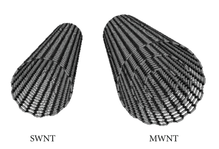
Carbon nanotubes (CNTs) are graphene sheets rolled into a cylindrical shape. Several sheets may roll into MWNTs whilst a single sheet rolls into a SWNT [2].
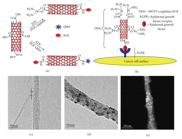
(a) Schematic representation of functionalization of SWNTs with quantum dots, EDC, and cisplatin; (b) SWNT bioconjugated with cisplatin and EGF, targeting cancer cell surface receptor EGFR; (c)–(e) TEM images showing the various functional groups, with cisplatin shown as bright spots [7].
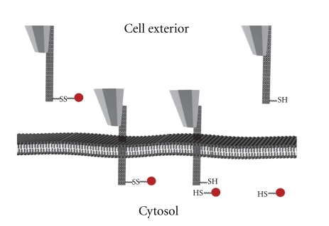
A schematic diagram showing that an AFM-controlled MWNT-based nanoinjector was able to penetrate into a cell and release the attached cargo compound after the breakage of the disulfide bond. This was followed by successful retraction of the nanoinjector with no apparent cell damage being produced [6].
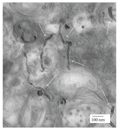
The perpendicular positioning of MWNTs (pointed at by the white arrows) during internalization into HeLa cells suggests that cellular uptake of CNTs by the cells was similar to that of nanoneedles [5].
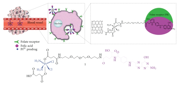
The “longboat” anticancer system in which the chemotherapeutic agent cisplatin is attached from one end to the FA derivative and from the opposite end to a SWNT via an amide link [20].
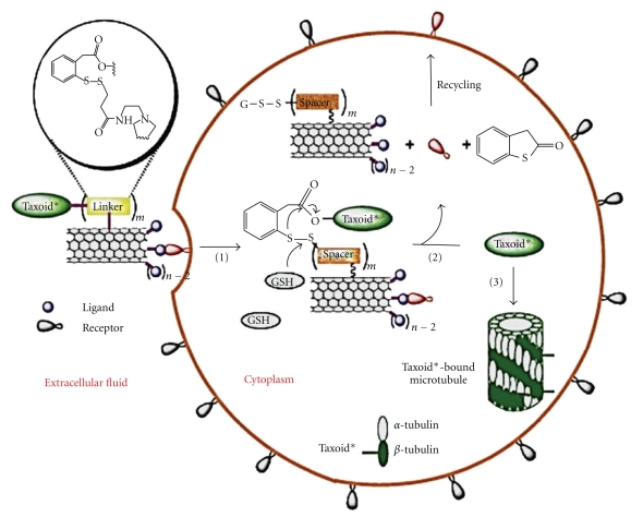
(1) Internalization of the CNTs carried conjugate into the tumour cell via receptor-mediated endocytosis. (2) Taxoid was released by the cleavage of the chemical linker. (3) The free taxoid molecules were bound to microtubules to form stabilized microtubules, resulting in arrest of cell mitosis and induction of apoptosis [31].
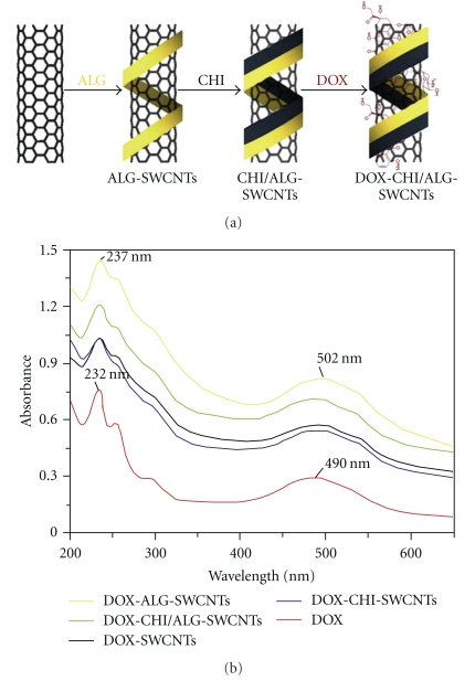
(a) Preparation of SWNTs-DOX after inclusion of bioadhesive polymers to enhance nanotubes dispersability in aqueous phase. (b) UV absorption spectra of DOX formulations [32].
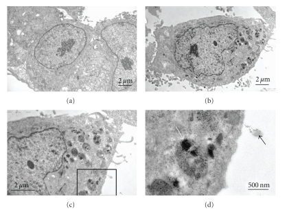
TEM showing the difference between HeLa cancer cells before treatment and after treatment with carbon nanotube formulations and the fate of the nanotubes: (a) HeLa cells before treatment, (b) HeLa cells treated with DOX-FA-CHI-ALG-SWNTs, (c) a magnified image of (b), and (d) magnified image of the boxed region in (c). The black arrow points at a SWNT-containing vesicle, and the white arrow points at some aggregated nanotubes inside a lysosome [32].
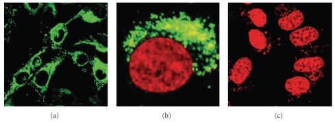
Confocal microscopy showing the internalization of labeled single strand DNA into Hela cell using SWNTs. (a) The labeled DNA (green colour) is surrounding the nucleus (black circles) at 37°C. (b) The nucleus stained using DRAQ5 (red color) is surrounded by the labeled DNA (green colour) after internalization at 37°C. (c) At 4°C, no DNA internalization has occurred [39].
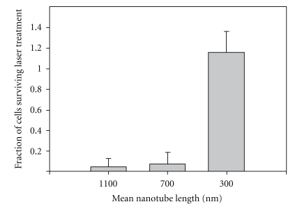
The relationship between cell survival and CNTs length using photothermal therapy. This has shown that nanotube lengths of 700 and 1100 nm are much more desirable in killing tumor cells compared with the length of 300 nm [57].
References
-
- Iijima S. Helical microtubules of graphitic carbon. Nature. 1991;354(6348):56–58.
-
- Pantarotto D, Briand JP, Prato M, Bianco A. Translocation of bioactive peptides across cell membranes by carbon nanotubes. Chemical Communications. 2004;10(1):16–17. - PubMed
-
- Pantarotto D, Singh R, McCarthy D, et al. Functionalized carbon nanotubes for plasmid DNA gene delivery. Angewandte Chemie International Edition. 2004;43(39):5242–5246. - PubMed
-
- Bianco A, Kostarelos K, Prato M. Applications of carbon nanotubes in drug delivery. Current Opinion in Chemical Biology. 2005;9(6):674–679. - PubMed
LinkOut - more resources
Full Text Sources
