. 2011 Oct 26:7:84.
doi: 10.1186/1744-8069-7-84.

Visceral hyperalgesia induced by forebrain-specific suppression of native Kv7/KCNQ/M-current in mice

Affiliations

Visceral hyperalgesia induced by forebrain-specific suppression of native Kv7/KCNQ/M-current in mice

Yeping Bi et al. Mol Pain. .

Abstract

Background: Dysfunction of brain-gut interaction is thought to underlie visceral hypersensitivity which causes unexplained abdominal pain syndromes. However, the mechanism by which alteration of brain function in the brain-gut axis influences the perception of visceral pain remains largely elusive. In this study we investigated whether altered brain activity can generate visceral hyperalgesia.

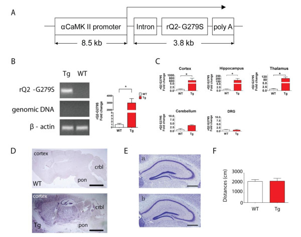
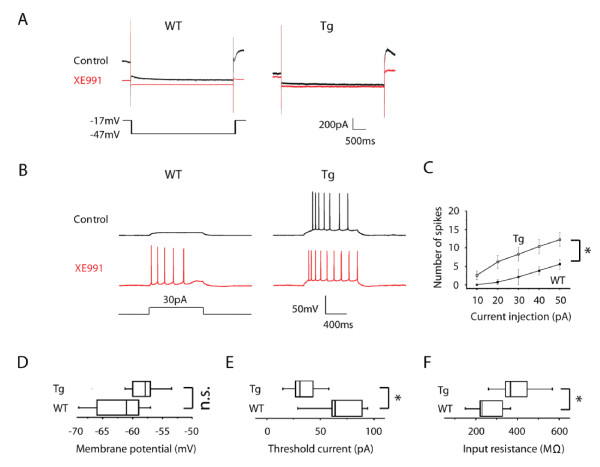
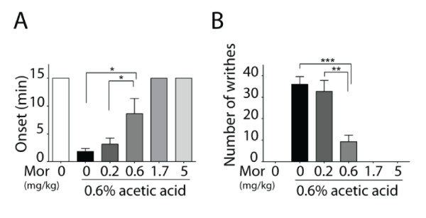
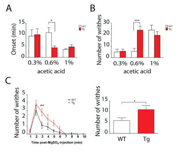
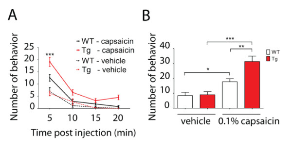
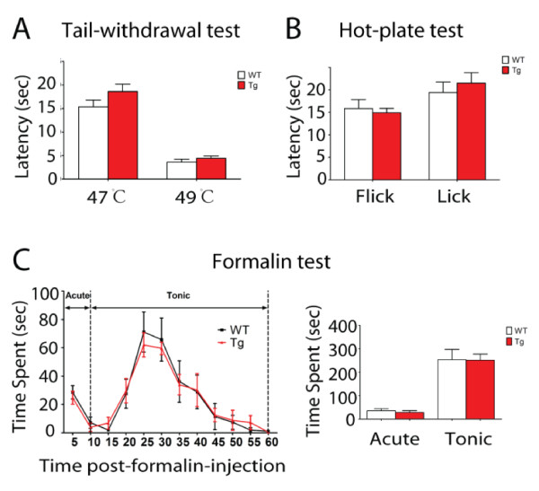
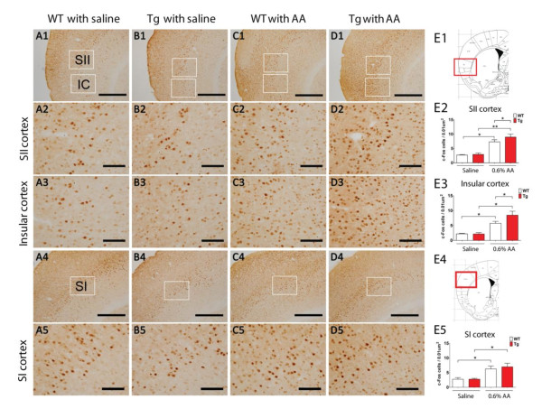
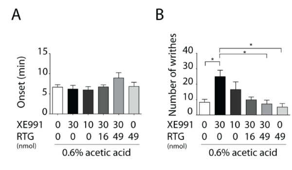
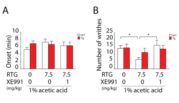
Results: Using a forebrain specific αCaMKII promoter, we established a line of transgenic (Tg) mice expressing a dominant-negative pore mutant of the Kv7.2/KCNQ2 channel which suppresses native KCNQ/M-current and enhances forebrain neuronal excitability. Brain slice recording of hippocampal pyramidal neurons from these Tg mice confirmed the presence of hyperexcitable properties with increased firing. Behavioral evaluation of Tg mice exhibited increased sensitivity to visceral pain induced by intraperitoneal (i.p.) injection of either acetic acid or magnesium sulfate, and intracolon capsaicin stimulation, but not cutaneous sensation for thermal or inflammatory pain. Immunohistological staining showed increased c-Fos expression in the somatosensory SII cortex and insular cortex of Tg mice that were injected intraperitoneally with acetic acid. To mimic the effect of cortical hyperexcitability on visceral hyperalgesia, we injected KCNQ/M channel blocker XE991 into the lateral ventricle of wild type (WT) mice. Intracerebroventricular injection of XE991 resulted in increased writhes of WT mice induced by acetic acid, and this effect was reversed by co-injection of the channel opener retigabine.

Conclusions: Our findings provide evidence that forebrain hyperexcitability confers visceral hyperalgesia, and suppression of central hyperexcitability by activation of KCNQ/M-channel function may provide a therapeutic potential for treatment of abdominal pain syndromes.


Figures

Figure 1
Figure 1
Generation and confirmation of transgenic mice overexpressing the KCNQ2 dominant-negative pore mutant in forebrain. A. Schematic organization of the transgene for forebrain-specific expression of the dominant-negative mutant of rat KCNQ2-G279S (rQ2-G279S) driven by the αCaMKII promoter. B. Identification of rQ2-G279S mRNA in the brain isolated from transgenic (Tg) and wild type (WT) mice by real time RT-PCR (mean ± s.e.m., n = 4, *p < 0.05). The template of rQ2-G279S specific primers for detection of KCNQ2 mutant gene expression is located in the area of SV40 poly A of the 265-plasmid (transgenic construct). The template of genomic DNA primers for detection of genomic DNA contamination is located after SV40 poly A in the 265-plasmid (transgenic construct), which does not undergo transcription. β-actin is used as an internal reference. C. Expression of rQ2-G279S mRNA isolated from 5 different tissues of WT and Tg mice by real time RT-PCR. The transgene rQ2-G279S was adequately expressed in the cortex, hippocampus and thalamus, with little expression in the cerebellum, and was not expressed in the DRG of Tg mice (mean ± s.e.m., n = 4, *p < 0.05). D. Expression of rQ2-G279S mRNA of WT (top) and Tg mice (down) using in situ hybridyzation. The transgene rQ2-G279S was adequately expressed in the cortex, hippocampus, and thalamus, with little expression in the cerebellum (crbl) and pons, and was not expressed in the medulla of Tg mice. The scale bar is 2mm. E. Nissl-stained coronal sections of hippocampus. No obvious structural change was found in adult transgenic mice (a) or wild type littermates (b). Scale bar, 500 μm. F. Locomotor activity test, no significant difference was detected in wild type (WT) and transgenic mice (Tg) in the total distance traveled during a period of 30 minutes (mean ± s.e.m., n = 7).
Figure 2
Figure 2
Electrophysiological characterization of CA1 pyramidal neurons from wild type and transgenic mice. A. Representative deactivation currents recorded from CA1 pyramidal neurons in fresh slice preparations from wild type (WT, left panel) or transgenic mice (Tg, right panel). Overlay of currents evoked by the protocol in the absence (black) or presence (red) of 10uM XE991. B. Response of CA1 pyramidal neurons with injection of 1000ms, 30pA depolarizing current pulse into the cell from WT (left panels) and Tg mice (right panels) before (black) and after (red) application of XE991. XE991 increased action potential firing in WT mice, but it was less effective in neurons from Tg mice. C. Comparison of average number of spikes evoked by injected current in WT and Tg neurons. D-F. Box plots illustrating the differences in WT and Tg mice in resting membrane potential, threshold current required for action potential firing and input resistance, n = 10; Data are shown as mean ± s.e.m., *p < 0.05.
Figure 3
Figure 3
Validation of visceral pain induced with acetic acid. Visceral pain was induced with acetic acid in ICR species of mice. The onset (A) and the total number of writhes (B) during a period of 15 minutes were determined after s.c. administration of morphine (0.2, 0.6, 1.7, 5 mg/kg) 20 minutes before i.p. injection of 0.6% acetic acid. Intraperitoneal injection of 0.6% AA resulted in a significant reduction in onset time of writhes and increased number of writhes. Morphine significantly increased the onset and decreased the total number of writhes at a dose of 0.6-5 mg/kg (mean ± s.e.m., n = 6-10, *p < 0.05, **p < 0.01, ***p < 0.001).
Figure 4
Figure 4
Visceral hyperalgesia in transgenic mice induced by visceral stimulation of acetic acid or magnesium sulfate. A-B. The onset (A) and the total number of writhes (B) during a period of 15 minutes were determined after i.p. injection of 0.3%, 0.6%, 1% acetic acid (n = 7-10). 0.6% acetic acid resulted in a shorted onset and increased number of writhes in transgenic (Tg) mice. C. Induction of visceral pain induced by magnesium sulfate (12 mg/k g, i.p.). Tg mice displayed an increase of writhes in response to magnesium sulfate during a period of 10 minutes (mean ± s.e.m., n = 10, *p < 0.05, **p < 0.01, ***p < 0.001).
Figure 5
Figure 5
Visceral hyperalgesia in transgenic mice induced by intracolon injection of capsaicin. Behavioral reactions (abdominal licking, stretching, squashing the lower abdomen against floor and abdominal retractions) evoked by intracolonic injection of vehicle solution or 0.1% capsaicin in WT and Tg mice. A. Number of behaviors observed in each 5 min period over a total observation time of 20 min post-administration. ***Indicates the other three groups which were significantly different from capsaicin-treated Tg mice during the first 5 min observation period (p < 0.001). B. Number of behaviors observed in the total 20 min period post-administration. Data are shown as mean ± s.e.m. (n = 5-11, *p < 0.05, **p < 0.01, ***p < 0.001).
Figure 6
Figure 6
Normal cutaneous pain sensitivity in transgenic mice to thermal and inflammatory stimulation. A-B. Comparison of thermal sensitivity in wild type (WT) and transgenic mice (Tg) by cutaneous application of thermal stimulation. No significant difference was detected between the two groups in tail withdrawal test (A) at 47°C and 49°C and in hot plate test (B) at 53°C (mean ± s.e.m, n = 7-11). C. The Tg mice displayed equivalent licking/biting behavior in both the acute and tonic phases in 5% formalin test (mean ± s.e.m, n = 7-9). The acute phase of the formalin test was defined as 0-10 min after injection and the tonic phase as 10-60 min after injection.
Figure 7
Figure 7
Induction of c-fos expression in forebrain cortex by 0.6% acetic acid stimulation. A-D. Representative photomicrographs of saline or acetic acid (i.p.) induced c-Fos expression in somatosensory SII cortex, insular cortex (IC) and SI cortex in wild type (WT) and transgenic (Tg) mice. The scale bar for panels A1-D1 and A4-D4 is 500 μm (low magnification), and the scale bar for A2-D2, A3-D3 and A5-D5 is 100 μm (high magnification). E1, Brain map [52] with photographed somatosensory SII and insular cortex framed in red. E2-3, Density of c-Fos positive cells in somatosensory SII cortex (E2) and Insular cortex (E3) of WT and Tg mice treated with saline or 0.6% acetic acid (i.p.). E4, Brain map [52] with the SI cortex framed in red. E5, Density of c-Fos positive cells in the SI cortex from WT and Tg mice treated with saline or 0.6% acetic acid (i.p.).Data are presented as mean ± s.e.m (n = 3-4, *p < 0.05, **p < 0.01).
Figure 8
Figure 8
Effect of intracerebroventricular (i.c.v.) administration of KCNQ channel modulators on visceral pain induced by acetic acid in wild type mice. The onset (A) and the total number of writhes (B) after induction of visceral pain by 0.6% acetic acid (i.p.) were determined over a period of 15 min with XE991, retigabine (RTG) or vehicle administered by i.c.v. injection. XE991 (30 nmol) significantly increased the number of writhes, and RTG (49 nmol) reversed its effect. Data are expressed as mean ± s.e.m (n = 7-13, *p < 0.05).
Figure 9
Figure 9
Antinociceptive effects of KCNQ channel opener retigabine on visceral pain induced by acetic acid in wild type mice, but not transgenic mice. The onset time (A) and the total number of writhes (B) were determined with vehicle or retigabine (RTG, 7.5 mg/kg) or RTG (7.5 mg/kg) co-injected with XE991 (1 mg/kg) administration (i.p.) before 1% acetic acid (i.p.) injection in both wild type (WT) and transgenic (Tg) groups. Data are expressed as mean ± s.e.m (n = 8-13). RTG (7.5 mg/kg, i.p.) significantly reduced the number of writhes in WT mice, and co-injection of RTG (7.5 mg/kg, i.p.) with XE991 (1 mg/kg, i.p.) reversed the antinociceptive effect of RTG (*p < 0.05).

Similar articles

Cited by

References

    1. Anand P, Aziz Q, Willert R, van Oudenhove L. Peripheral and central mechanisms of visceral sensitization in man. Neurogastroenterol Motil. 2007;19:29–46. doi: 10.1111/j.1365-2982.2006.00873.x. - DOI - PubMed
    1. Woolf CJ. Central sensitization: implications for the diagnosis and treatment of pain. Pain. 2011;152:S2–15. doi: 10.1016/j.pain.2010.09.030. - DOI - PMC - PubMed
    1. Latremoliere A, Woolf CJ. Central sensitization: a generator of pain hypersensitivity by central neural plasticity. J Pain. 2009;10:895–926. doi: 10.1016/j.jpain.2009.06.012. - DOI - PMC - PubMed
    1. Mayer EA, Tillisch K. The brain-gut axis in abdominal pain syndromes. Annu Rev Med. 2011;62:381–396. doi: 10.1146/annurev-med-012309-103958. - DOI - PMC - PubMed
    1. Moisset X, Bouhassira D, Denis D, Dominique G, Benoit C, Sabate JM. Anatomical connections between brain areas activated during rectal distension in healthy volunteers: a visceral pain network. Eur J Pain. 2010;14:142–148. doi: 10.1016/j.ejpain.2009.04.011. - DOI - PubMed

Publication types

MeSH terms

LinkOut - more resources