Comparative Study
. 2011;13(5):R179.
doi: 10.1186/ar3504. Epub 2011 Oct 26.

Impact of interleukin-21 in the pathogenesis of primary Sjögren's syndrome: increased serum levels of interleukin-21 and its expression in the labial salivary glands

Affiliations
Comparative Study

Impact of interleukin-21 in the pathogenesis of primary Sjögren's syndrome: increased serum levels of interleukin-21 and its expression in the labial salivary glands

Kwi Young Kang et al. Arthritis Res Ther. 2011.

Abstract

Introduction: Interleukin (IL)-21 is a cytokine that controls the functional activity of effector T helper cells and the differentiation of Th17 cells, and promotes B-cell differentiation. To test whether IL-21 participates in the pathogenesis of primary Sjögren's syndrome (SS), serum IL-21 level was measured and IL-21 expression in the labial salivary glands (LSG) was examined.

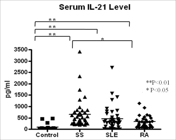
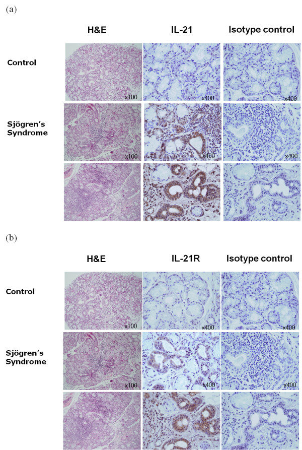
Methods: Serum IL-21 levels in 40 primary SS, 40 rheumatoid arthritis (RA), and 38 systemic lupus erythematosus (SLE) patients and 20 healthy controls were measured. Serum IL-21 levels of SS patients were assessed for correlations with laboratory data, including anti-nuclear antibody, anti-Ro/La antibodies, globulin, immunoglobulin (Ig) class, and IgG subclass. LSGs from 16 primary SS and 4 controls with sicca symptoms were evaluated for IL-21 and IL-21 receptor (IL-21R) expression by immunohistochemistry. Confocal microscopy was performed to further characterize the IL-21 positive cells.

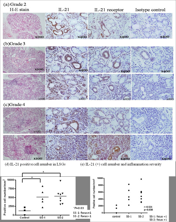
Results: Primary SS patients had significantly higher serum IL-21 levels than controls, and these increments correlated positively with levels of IgG, IgG1. Serum IgG1 levels correlated with anti-Ro antibody titers. Immunohistochemical analyses showed that lymphocytic foci and the periductal area of the LSGs from SS patients expressed high levels of IL-21 and lower levels of IL-21R, whereas the control LSGs showed minimal expression of both antigens. The more the lymphocyte infiltrated, IL-21 expression in LSGs showed a tendency to increase. Confocal microscopic analyses revealed that IL-21 expressing infiltrating lymphocytes in the LSGs of SS patients also expressed CXCR5.

Conclusions: Primary SS is associated with high serum IL-21 levels that correlate positively with serum IgG, especially IgG1, levels. The expression of IL-21 is increased as more lymphocytes infiltrated in LSGs. These observations suggest that IL-21 may play an important role in primary SS pathogenesis.


Figures

Figure 1
Figure 1
The serum IL-21 level. The serum IL-21 levels of patients with primary SS (n = 40), SLE (n = 38), or RA (n = 40) and healthy controls (n = 20) were measured by ELISA. The primary SS patients had significantly higher serum IL-21 levels than the healthy controls or the RA patients. Horizontal bars represent the median of all samples within a group. * P < 0.05, ** P < 0.01. IL: Interleukin; RA: rheumatoid arthritis; SLE: systemic lupus erythematosus; SS: Sjögren's syndrome.
Figure 2
Figure 2
Correlations between the serum IL-21 levels of primary SS patients and the levels of immunoglobulin. (a) The serum IL-21 levels of primary SS patients (n = 40) correlated significantly with the serum globulin levels. (b) Of all the Ig classes, the serum IL-21 levels only correlated with IgG (n = 40). (c) There was a positive correlation between the serum IL-21 and IgG1 levels (n = 24). (d) The anti-Ro/SSA antibody titers, as measured by ELISA, correlated with the IgG1 levels (n = 35). (e) The serum IL-21 levels correlated with anti-Ro/SSA antibody index (n = 24) and IL-21 was an independent variable in regression analysis. The correlation coefficients (r) and P-alues of statistical significance are shown. IG: immunoglobulin; IL: Interleukin; SS: Sjögren's syndrome.
Figure 3
Figure 3
The labial salivary glands (LSG) of primary SS exhibit increased IL-21 and IL-21 receptor expression. Shown is the expression of the IL-21 and IL-21 receptor in the labial salivary glands of control subjects (n = 4; upper panel) and patients with primary SS (n = 16; lower two panels), as determined by immunostaining using specific antibodies. The cells that stained with the antibodies appeared in brown. (a) The infiltrating lymphocytes and periductal areas of the patients with SS exhibited intense IL-21 staining, whereas there was no staining with the isotype controls. (b) The lymphocytic infiltrations and periductal areas stained for IL-21 receptor. In contrast, the control subjects did not exhibit any IL-21 or IL-21 receptor expression in their labial salivary glands. IL: Interleukin; LSGs: labial salivary glands; SS: Sjögren's syndrome.
Figure 4
Figure 4
The expression of IL-21 in the LSG of SS patients and the severity of inflammation. The labial salivary gland specimens of the primary SS patients were divided according to the grade (1 to 4) of lymphocytic infiltration (see Methods). (a) This specimen was obtained from a SS patient who fulfilled the American-European Consensus Group Criteria except LSG biopsy. There were no definite lymphocytic foci but moderate lymphocytic infiltration was observed. (a-c) IL-21 expression increased as the lymphocytic infiltration in the salivary gland became more severe. (d) IL-21 positive cell number was significantly increased in LSG from SS patients than in control. (e) Relationship between IL-21 positive cell number and inflammation severity. IL-21 positive cell number was elavated with grade (r = 0.521, P = 0.038). Horizontal bars represent the median of all samples within a group. Representative data are shown. IL-21: Interleukin-21; LSGs: labial salivary glands; SS: Sjögren's syndrome.
Figure 5
Figure 5
Confocal microscopic analysis of the IL-21-expressing cells in the LSGs of primary SS patients. (a) Immunofluorescent double-staining of IL-21-expressing cells (red) and CXCR5+ cells (blue). (b) Immunofluorescent double-staining of CD4+ T cells (green) and CXCR5+ cells (blue). (c) Immunofluorescent double-staining of IL-21-expressing cells (red) and CD20+ B cells (green). Only the IL-21-expressing cells merged with the CXCR5+ cells. IL-21: Interleukin-21; LSGs: labial salivary glands; SS: Sjögren's syndrome.

Comment in

  • IL-21 and Sjögren's syndrome.
    Scofield RH. Scofield RH. Arthritis Res Ther. 2011;13(6):137. doi: 10.1186/ar3518. Epub 2011 Dec 19. Arthritis Res Ther. 2011. PMID: 22226370 Free PMC article.

References

    1. Spolski R, Leonard WJ. Interleukin-21: basic biology and implications for cancer and autoimmunity. Annu Rev Immunol. 2008;26:57–79. doi: 10.1146/annurev.immunol.26.021607.090316. - DOI - PubMed
    1. Coquet JM, Kyparissoudis K, Pellicci DG, Besra G, Berzins SP, Smyth MJ, Godfrey DI. IL-21 is produced by NKT cells and modulates NKT cell activation and cytokine production. J Immunol. 2007;178:2827–2834. - PubMed
    1. Ozaki K, Spolski R, Feng CG, Qi CF, Cheng J, Sher A, Morse HC, Liu C, Schwartzberg PL, Leonard WJ. A critical role for IL-21 in regulating immunoglobulin production. Science. 2002;298:1630–1634. doi: 10.1126/science.1077002. - DOI - PubMed
    1. Monteleone G, Pallone F, Macdonald TT. Interleukin-21 (IL-21)-mediated pathways in T cell-mediated disease. Cytokine Growth Factor Rev. 2009;20:185–191. doi: 10.1016/j.cytogfr.2009.02.002. - DOI - PubMed
    1. Ettinger R, Kuchen S, Lipsky PE. Interleukin 21 as a target of intervention in autoimmune disease. Ann Rheum Dis. 2008;67(Suppl 3):iii83–86. doi: 10.1136/ard.2008.098400. - DOI - PubMed

Publication types