. 2012 Jan 15;71(2):120-8.
doi: 10.1016/j.biopsych.2011.09.011. Epub 2011 Oct 25.

Nicotinic α5 subunits drive developmental changes in the activation and morphology of prefrontal cortex layer VI neurons

Affiliations

Nicotinic α5 subunits drive developmental changes in the activation and morphology of prefrontal cortex layer VI neurons

Craig D C Bailey et al. Biol Psychiatry. .

Abstract

Background: Nicotinic signaling in prefrontal layer VI pyramidal neurons is important to the function of mature attention systems. The normal incorporation of α5 subunits into α4β2* nicotinic acetylcholine receptors augments nicotinic signaling in these neurons and is required for normal attention performance in adult mice. However, the role of α5 subunits in the development of the prefrontal cortex is not known.

Methods: We sought to answer this question by examining nicotinic currents and neuronal morphology in layer VI neurons of medial prefrontal cortex of wild-type and α5 subunit knockout (α5(-/-)) mice during postnatal development and in adulthood.

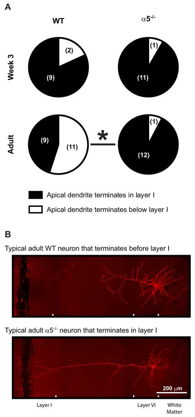
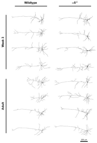
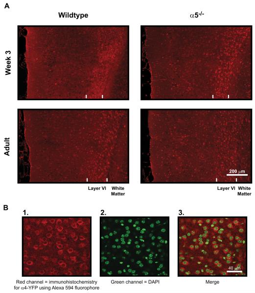
Results: In wild-type but not in α5(-/-) mice, there is a developmental peak in nicotinic acetylcholine currents in the third postnatal week. At this juvenile time period, the majority of neurons in all mice have long apical dendrites extending into cortical layer I. Yet, by early adulthood, wild-type but not α5(-/-) mice show a pronounced shift toward shorter apical dendrites. This cellular difference occurs in the absence of genotype differences in overall cortical morphology.

Conclusions: Normal developmental changes in nicotinic signaling and dendritic morphology in prefrontal cortex depend on α5-comprising nicotinic acetylcholine receptors. It appears that these receptors mediate a specific developmental retraction of apical dendrites in layer VI neurons. This finding provides novel insight into the cellular mechanisms underlying the known attention deficits in α5(-/-) mice and potentially also into the pathophysiology of developmental neuropsychiatric disorders such as attention-deficit disorder and autism.


Figures

Figure 1
Figure 1
Changes in the excitation of medial prefrontal layer VI pyramidal neurons by acetylcholine during postnatal development are dependent on the nicotinic receptor α5 subunit. (A) The peak inward current response to 10-second bath application of 1 mmol/L acetylcholine (in the presence of 200 nmol/L atropine) was measured in neurons from wild-type (WT) mice expressing the α5 subunit (closed bars) and in neurons from mice in which the α5 subunit has been genetically deleted (α5−/−, open bars) in four postnatal age groups (week 2, week 3, week 4, and adult). Two-way analysis of variance identified an effect of genotype [F(1,166) = 80.72, p < .0001], where the current response in WT neurons was greater than that in α5−/− neurons at each age (Bonferroni post hoc test, *p < .05 at each age), and also a significant interaction between the effects of genotype and age [F(3,166) = 2.98, p = .03]. There was a significant effect of age in WT neurons only, where the acetylcholine response at week 3 was significantly greater than each of the other ages [one-way analysis of variance, F(3,86) = 2.92, p = .04; Newman-Keuls post hoc test, p < .05 compared with each other age]. Typical voltage-clamp traces in response to acetylcholine application (dark gray lines) are shown in (B) for neurons from each experimental group.
Figure 2
Figure 2
Qualitative analysis of medial prefrontal layer VI pyramidal neuron apical dendrites. Pie charts in (A) show the proportion of neurons from wild-type (WT) mice and nicotinic receptor α5 subunit knockout (α5−/−) mice at postnatal week 3 and in adulthood with apical dendrite trees that either terminate within layer I (black portion) or terminate below layer I (white portion) of the medial prefrontal cortex. The absolute number of neurons identified within each group is shown in brackets. At week 3, the majority of neurons of both genotypes had long apical dendrites extending into layer I, whereas in adulthood, the majority of WT neurons had shorter apical dendrites that terminated below layer I (Fisher’s exact test, p = .07 comparing adult WT and week 3 WT neurons; *p = .009 comparing adult WT and adult α5−/− neurons). Representative z-projection photomicrographs of Neurobiotin-filled neurons are shown in (B) for a typical adult WT neuron that terminates below layer I and a typical adult α5−/− neuron that terminates within layer I.
Figure 3
Figure 3
Representative z-projections of traced neurons are shown for wild-type and nicotinic receptor α5 subunit knockout (α5−/−) mice at postnatal week 3 and in adulthood.
Figure 4
Figure 4
Quantitative analysis of medial prefrontal layer VI pyramidal neuron apical dendrites. (A–D) Three-dimensional Sholl analysis measuring the number of dendrite intersections at concentric spheres of varying distance from the soma for neurons from wild-type (WT) and nicotinic receptor α5 subunit knockout (α5−/−) mice. Two-way analysis of variance found effects of genotype both at week 3 (A) [F(1,578) = 5.97, p = .01] and in adulthood (B) [F(1,1044) = 40.88, p < .0001]. Comparison of the Sholl analyses for WT mice at each age (C) identified a significant effect of age [F(1,850) = 6.65, p = .01] with the difference between ages appearing at the most distal 300 μm from the soma (marked by an arrow), whereas the same comparison in α5−/− mice (D) found no effect of age [F(1,756) = 0.3, p = .6]. Two-way analysis of variance identified significant effects of α5 subunit genotype on total apical dendrite length (E) [F(1,46) = 5.81, p = .02], the distance from the soma to the most distal apical dendrite terminal (F) [F(1,46) = 5.15, p = .03], and the total number of apical dendrite terminals (G) [F(1,46) = 4.85, p = .03]. However, there were no significant effects of age or significant interactions between the effects of genotype and age on these three measures (all p > .05). *p < .05 for the difference between WT and α5−/− neurons in adulthood (Bonferroni post hoc test).
Figure 5
Figure 5
Immunohistochemistry for α4* nicotinic acetylcholine receptors within the medial prefrontal cortex. (A) Low magnification photomicrographs within medial prefrontal slices showing immunohistochemical staining for nicotinic receptor α4 subunits tagged with the yellow fluorescent protein (YFP) motif in wild-type and nicotinic receptor α5 subunit knockout (α5−/−) mice at postnatal week 3 and in adulthood. The pial (medial) surface is located on the left and the white matter is located on the right of each photomicrograph. Immunostaining in all mice identified a distinct band of bright red neuronal cell bodies expressing α4* nicotinic receptors within layer VI (presumably α4β2* receptors). This band was thinner and more dense at week 3 compared with adulthood. However, there were no effects of α5 subunit genotype on the width of the layer VI immunoreactive band, the proportion of neurons within the band expressing α4* nicotinic receptors, or the width of the medial prefrontal cortex (see Results for more detail). (B) Example high-resolution photomicro-graphs are shown within layer VI for 1) α4-YFP immunostained neurons, 2) 4 ,6-diamidino-2-phenylindole (DAPI) counterstained cell nuclei, and 3) the merge of α4-YPP and DAPI staining from 1) and 2).

References

    1. Passetti F, Dalley JW, O’Connell MT, Everitt BJ, Robbins TW. Increased acetylcholine release in the rat medial prefrontal cortex during performance of a visual attentional task. Eur J Neurosci. 2000;12:3051–3058. - PubMed
    1. Dalley JW, Theobald DE, Bouger P, Chudasama Y, Cardinal RN, Robbins TW. Cortical cholinergic function and deficits in visual attentional performance in rats following 192 IgG-saporin-induced lesions of the medial prefrontal cortex. Cereb Cortex. 2004;14:922–932. - PubMed
    1. Parikh V, Kozak R, Martinez V, Sarter M. Prefrontal acetylcholine release controls cue detection on multiple timescales. Neuron. 2007;56:141–154. - PMC - PubMed
    1. Kassam SM, Herman PM, Goodfellow NM, Alves NC, Lambe EK. Developmental excitation of corticothalamic neurons by nicotinic acetylcholine receptors. J Neurosci. 2008;28:8756–8764. - PMC - PubMed
    1. Alves NC, Bailey CDC, Nashmi R, Lambe EK. Developmental sex differences in nicotinic currents of prefrontal layer VI neurons in mice and rats. PLoS One. 2010;5:e9261. - PMC - PubMed

Publication types

LinkOut - more resources