

Hemangiogenesis and lymphangiogenesis in corneal pathology
- PMID: 22030600
- PMCID: PMC3710690
- DOI: 10.1097/ICO.0b013e31821dd986
Hemangiogenesis and lymphangiogenesis in corneal pathology
Abstract
Purpose: We characterized the presence of hemangiogenesis (HA) and lymphangiogenesis (LA) in human corneal specimens exhibiting 13 underlying pathologies.
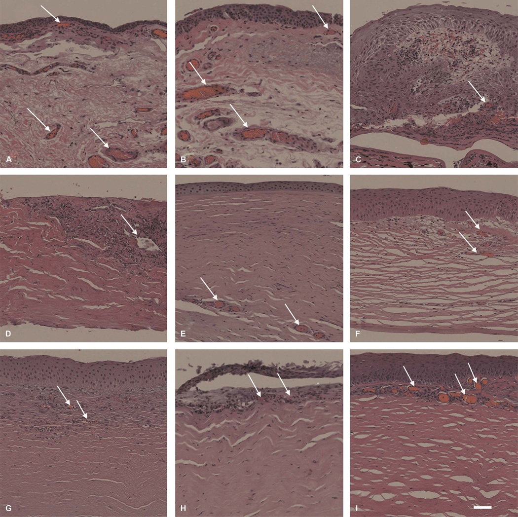
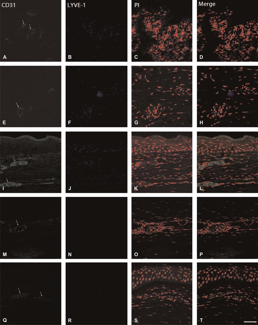
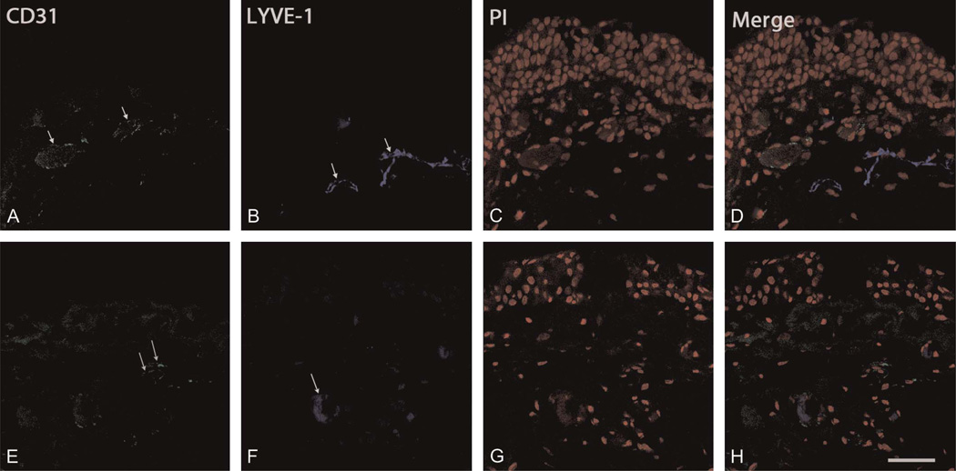
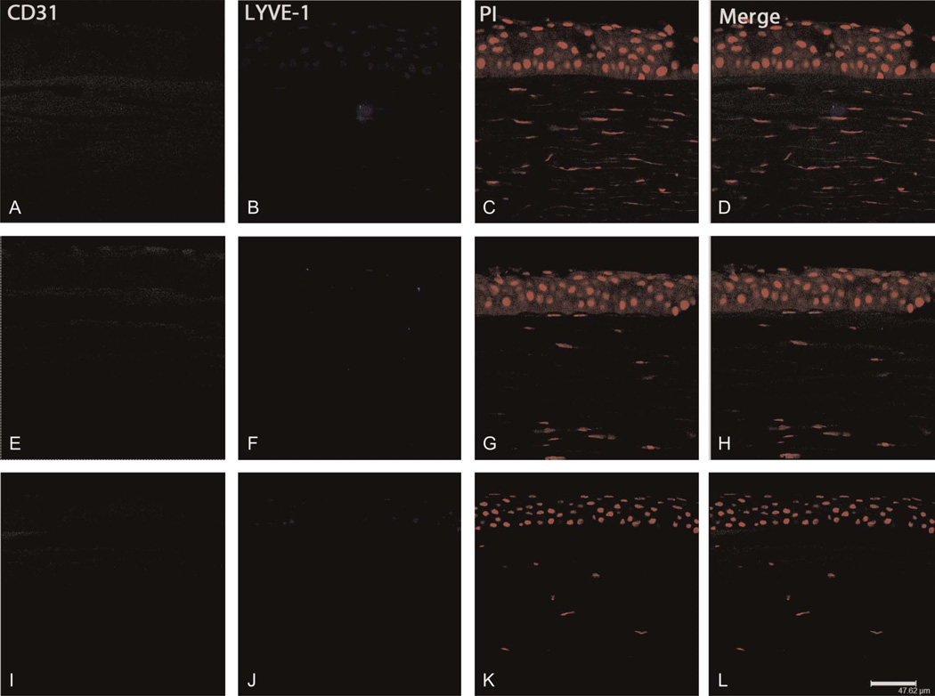
Methods: Human corneal specimens were obtained from consenting subjects (n = 2 or n = 3 for each pathology; total sample size, n = 35). The pathological specimens were stained with hematoxylin and eosin (H&E) to determine the presence or absence of corneal neovascularization (NV) and superficial or deep stromal distribution of NV. Immunohistochemical staining was then performed to differentiate HA (positive for CD31) from LA (positive for lymphatic vessel endothelial hyaluronan receptor-1 [LYVE-1]).
Results: The double-negative (CD31(-)/LYVE-1(-)) immunostaining, indicating the absence of NV, was exhibited by 21 specimens (60%). CD31(-)/LYVE-1(-), indicating the presence of HA and absence of LA, was exhibited by 12 specimens (34%). The double-positive (CD31(+)/LYVE-1(+)) phenotype, indicating both HA and LA, was exhibited by 2 specimens (6%). Notably, the CD31(-)/LYVE-1(-) phenotype, indicating the presence of LA and absence of HA, was not detected among the specimens. Deep stromal NV was exhibited in a 4:3 ratio to superficial stromal NV. The double-negative immunostaining was more prevalent in noninflammatory pathologies, particularly in comparison with combined neovascular phenotypes (ie, CD31(+) or LYVE-1(+)). Among the neovascular phenotypes, HA was 7 times more common than LA. Specimens exhibiting LA presented only with the double-positive phenotype.
Conclusions: HA is the predominant component of NV in corneal pathologies. LA accompanies HA; however, isolated LA (from lymphatics in the conjunctiva) does not occur in these corneal pathologies. Our results suggest the potential therapeutic utility of targeting antineovascular therapies specifically for corneal HA and/or LA pathology.
Conflict of interest statement
The authors state that they have no financial or conflicts of interest to disclose.
Figures
References
-
- Cursiefen C, Maruyama K, Jackson DG, et al. Time course of angiogenesis and lymphangiogenesis after brief corneal inflammation. Cornea. 2006;25:443–447. - PubMed
-
- Regenfuss B, Bock F, Parthasarathy A, et al. Corneal (lymph)angiogenesis—from bedside to bench and back: a tribute to Judah Folkman. Lymphat Res Biol. 2008;6:191–201. - PubMed
-
- Chang JH, Gabison EE, Kato T, et al. Corneal neovascularization. Curr Opin Ophthalmol. 2001;12:242–249. - PubMed
Publication types
MeSH terms
Substances
Grants and funding
LinkOut - more resources
Full Text Sources
Miscellaneous
