. 2012 Jan;27(1):38-46.
doi: 10.1002/jbmr.516.

Longitudinal evaluation of FGF23 changes and mineral metabolism abnormalities in a mouse model of chronic kidney disease

Affiliations

Longitudinal evaluation of FGF23 changes and mineral metabolism abnormalities in a mouse model of chronic kidney disease

Jason R Stubbs et al. J Bone Miner Res. 2012 Jan.

Abstract

Fibroblast growth factor 23 (FGF23) is a phosphaturic and vitamin D-regulatory hormone of putative bone origin that is elevated in patients with chronic kidney disease (CKD). The mechanisms responsible for elevations of FGF23 and its role in the pathogenesis of chronic kidney disease-mineral bone disorder (CKD-MBD) remain uncertain. We investigated the association between FGF23 serum levels and kidney disease progression, as well as the phenotypic features of CKD-MBD in a Col4a3 null mouse model of human autosomal-recessive Alport syndrome. These mice exhibited progressive renal failure, declining 1,25(OH)(2)D levels, increments in parathyroid hormone (PTH) and FGF23, late-onset hypocalcemia and hyperphosphatemia, high-turnover bone disease, and increased mortality. Serum levels of FGF23 increased in the earliest stages of renal damage, before elevations in blood urea nitrogen (BUN) and creatinine. FGF23 gene transcription in bone, however, did not increase until late-stage kidney disease, when serum FGF23 levels were exponentially elevated. Further evaluation of bone revealed trabecular osteocytes to be the primary cell source for FGF23 production in late-stage disease. Changes in FGF23 mirrored the rise in serum PTH and the decline in circulating 1,25(OH)(2)D. The rise in PTH and FGF23 in Col4a3 null mice coincided with an increase in the urinary fractional excretion of phosphorus and a progressive decline in sodium-phosphate cotransporter gene expression in the kidney. Our findings suggest elevations of FGF23 in CKD to be an early marker of renal injury that increases before BUN and serum creatinine. An increased production of FGF23 by bone may not be responsible for early increments in FGF23 in CKD but does appear to contribute to FGF23 levels in late-stage disease. Elevations in FGF23 and PTH coincide with an increase in urinary phosphate excretion that likely prevents the early onset of hyperphosphatemia in the face of increased bone turnover and a progressive decline in functional renal mass.


Figures

Figure 1
Figure 1
Genotype (A), body weight (B), and survival (C) of Col4a3 wild-type and null mice (P<0.05 at 12 weeks for body weight and 14 weeks for survival, n ≥ 10 per group).
Figure 2
Figure 2
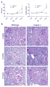
Evidence of progressive kidney disease in Col4a3 null mice. (A) Age-dependent changes in BUN (left panel) and serum creatinine (right panel) in wild-type and Col4a3null mice (# P<0.01, n ≥ 5 at each time point), (B) PAS staining of kidneys from 4, 6, and 10 week-old wild-type and Col4a3 null mice. Col4a3 null mice demonstrate normal renal histology at 4 weeks-of-age (top right panel), whereas the onset of significant tubular dilation/damage (thick arrows) and an interstitial infiltrate (arrowheads) are apparent by 6 weeks (middle right panel). After 10 weeks-of-age (bottom right panel), Col4a3 null mice exhibit significant glomerulosclerosis with crescent formation (thin arrow) and severe interstitial fibrosis (asterisks).
Figure 3
Figure 3
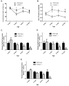
Evaluation of renal phosphorus excretion in Col4a3 null mice. Age-dependent changes in (A) fractional excretion of phosphorus and (B) total urine phosphorus were assessed in Col4a3 null and wild-type mice. Col4a3 null mice demonstrated an increase in both the fractional excretion of phosphorus and total urine phosphorus compared to wild-type mice beginning at 6 weeks. (C–E) Quantitative real-time PCR analysis of gene expression changes of NaPi-2a, NaPi-2c, and klotho in kidneys from Col4a3 null mice at 4 separate time points (4, 6, 8 and 12 weeks of age) were compared to those of wild-type littermates (wild-type gene expression was standardized to equal 1 for all time points). Col4a3 null mice demonstrated a progressive decline in NaPi-2a, NaPi-2c, and klotho gene expression between 6 and 12 weeks-of-age. (n ≥ 5 per group at each time point, * = P<0.05, ** = P<0.01, # = P<0.001 compared to wild-type mice of same age).
Figure 4
Figure 4
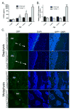
Serum changes of FGF23 relative to FGF23 gene expression changes in bone in Col4a3 null and wild-type mice. (A) Serum FGF23 levels at 4, 6, 8 & 12 weeks-of-age in Col4a3 null and wild-type mice. A progressive rise in circulating FGF23 levels begins around 6 weeks in null mice. (B) Quantitative real-time PCR analysis of FGF23 gene expression in calvaria from 4, 6, 8 & 12 week-old Col4a3 wild-type and null mice. No apparent difference in FGF23 gene expression at 4, 6, or 8 weeks in null mice, but a significant increase in calvarial expression of FGF23 is evident at 12 weeks (** = P<0.01, n ≥ 3 per group). (C) eGFP expression in distal femurs from 12 week-old Col4a3 wild-type and null mice that were crossed with eGFP reporter mice exhibiting eGFP expression (bright green fluorescence) under the control of the FGF23 promoter. Col4a3 null mice demonstrate no significant FGF23 expression by cortical osteocytes within the diaphysis (top panel), while trabecular osteocytes within the metaphysis (bright green cells in third panel) demonstrated significant FGF23 expression. All slides are presented at 20x magnification. Abbreviations: C – cortical bone, T – trabeculae, BM – bone marrow.
Figure 5
Figure 5
Col4a3 null mice develop high-turnover bone disease. (A) TRAP-stained sagittal section of long bone demonstrates increased osteoclast-mediated bone resorption in Col4a3 null mice (arrowheads in right inset identify red-staining osteoclasts). (B) Bone histomorphometry measurements of femurs from 10 week-old Col4a3 null and wild-type mice. Col4a3 null mice demonstrate a significant increase in osteoclast numbers (N.Oc/BS), and non-significant increases in both bone volume (BV/TV) and trabecular number (Tb.N) compared to wild-type mice. Abbreviations: trabecular thickness (Tb.Th), trabecular separation (Tb.Sp), osteoid thickness (O.Th), osteoid volume (OV/BV). (* = P<0.05, n ≥ 5 per group).

References

    1. Moe S, Drueke T, Cunningham J, et al. Definition, evaluation, and classification of renal osteodystrophy: a position statement from Kidney Disease: Improving Global Outcomes (KDIGO) Kidney Int. 2006;69:1945–53. - PubMed
    1. Gutierrez OM, Januzzi JL, Isakova T, et al. Fibroblast growth factor 23 and left ventricular hypertrophy in chronic kidney disease. Circulation. 2009;119:2545–52. - PMC - PubMed
    1. Gutierrez OM, Mannstadt M, Isakova T, et al. Fibroblast growth factor 23 and mortality among patients undergoing hemodialysis. N Engl J Med. 2008;359:584–92. - PMC - PubMed
    1. Mirza MA, Larsson A, Melhus H, Lind L, Larsson TE. Serum intact FGF23 associate with left ventricular mass, hypertrophy and geometry in an elderly population. Atherosclerosis. 2009;207:546–51. - PubMed
    1. Gutierrez O, Isakova T, Rhee E, et al. Fibroblast growth factor-23 mitigates hyperphosphatemia but accentuates calcitriol deficiency in chronic kidney disease. J Am Soc Nephrol. 2005;16:2205–15. - PubMed

Publication types

MeSH terms