

Inflammation of the fetal ovine skin following in utero exposure to Ureaplasma parvum
- PMID: 22031190
- PMCID: PMC3343146
- DOI: 10.1177/1933719111408114
Inflammation of the fetal ovine skin following in utero exposure to Ureaplasma parvum
Abstract
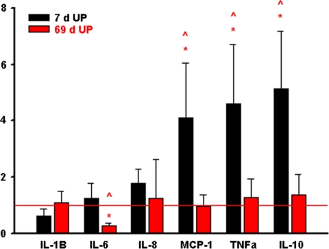
There is increasing evidence linking in utero infection and inflammation to preterm birth. Many commensal urogenital tract microorganisms, including the Mycoplasmas and Ureaplasmas, are commonly detected in association with preterm birth. Using an ovine model of sterile fetal inflammation, we demonstrated previously that the fetal skin generates a robust inflammatory response following in utero exposure to lipopolysaccharides from Escherichia coli. The fetal skin's response to colonization of the amniotic fluid by viable microorganisms remains unstudied. We hypothesised that in utero infection with Ureaplasma parvum serovar 3 would induce a proinflammatory response in the fetal skin. We found that (1) cultured fetal keratinocytes (the primary cellular constituent of the epidermis) respond to U. parvum exposure in vitro by increasing the expression of the chemotactant monocyte chemoattractant protein 1 (MCP-1) but not interleukin 1β (IL-1β), IL-6, IL-8, or tumor necrosis factor-α (TNF-α); (2) the fetal skin's response to 7 days of U. parvum exposure is characterized by elevated expression of MCP-1, TNF-α, and IL-10; and (3) the magnitude of inflammatory cytokine/chemokine expression in the fetal skin is dependent on the duration of U parvum exposure. These novel findings provide further support for the role of the fetal skin in the development of fetal inflammation and the preterm birth that may follow.
Conflict of interest statement
The authors declared no conflicts of interest with respect to the authorship and/or publication of this article.
Figures


References
-
- Janeway CA, Travers P, Walport M, Shlomchik M. Basic Concepts in Immunobiology. New York: Garland; 2001
-
- Liggins GC. Cervical ripening as an inflammatory reaction. In: DA Ellwood, ABM Anderson. (Eds.), The Cervix in Pregnancy and Labour, Clinical and Biochemical Investigations. Edinburgh: Churchill Livingstone; 1981:1–9
-
- Sennstromm MB, Ekman G, Westergren-Thorsson G, et al. Human cervical ripening, an inflammatory process mediated by cytokines. Mol Hum Reprod. 2000;6(4):375–381 - PubMed
-
- Kramer BW, Joshi SN, Moss TJ, et al. Endotoxin-induced maturation of monocytes in preterm fetal sheep lung. Am J Physiol Lung Cell Mol Physiol. 2007;293(2):L345–L353 - PubMed
Publication types
MeSH terms
Substances
Grants and funding
LinkOut - more resources
Full Text Sources
Research Materials
Miscellaneous
