. 2012 Mar;55(3):846-55.
doi: 10.1002/hep.24757.

Plasma cells and the chronic nonsuppurative destructive cholangitis of primary biliary cirrhosis

Affiliations

Plasma cells and the chronic nonsuppurative destructive cholangitis of primary biliary cirrhosis

Toru Takahashi et al. Hepatology. 2012 Mar.

Abstract

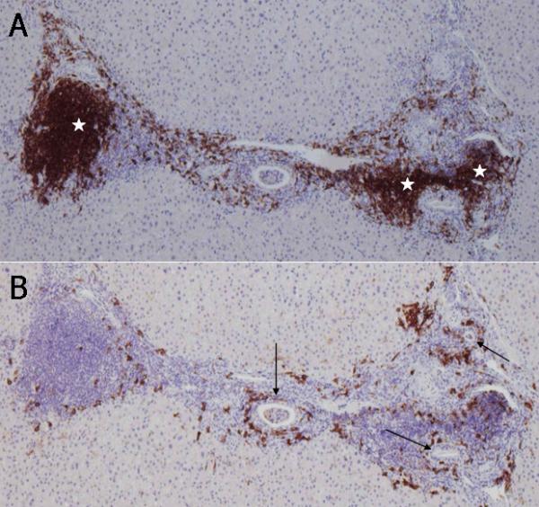
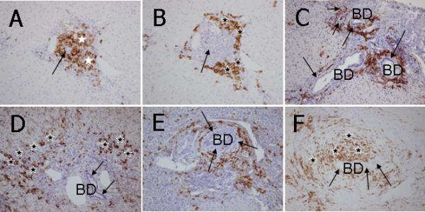
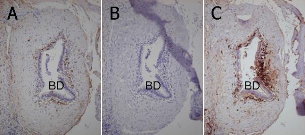
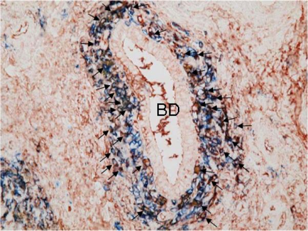
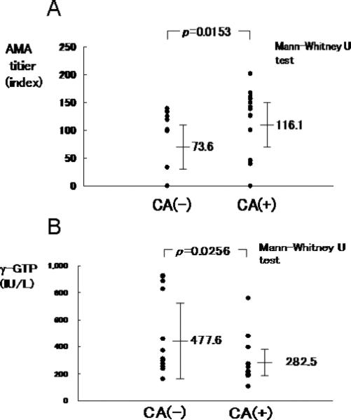
There has been increased interest in the role of B cells in the pathogenesis of primary biliary cirrhosis (PBC). Although the vast majority of patients with this disease have anti-mitochondrial antibodies, there is no correlation of anti-mitochondrial antibody titer and/or presence with disease severity. Furthermore, in murine models of PBC, it has been suggested that depletion of B cells may exacerbate biliary pathology. To address this issue, we focused on a detailed phenotypic characterization of mononuclear cell infiltrates surrounding the intrahepatic bile ducts of patients with PBC, primary sclerosing cholangitis, autoimmune hepatitis, chronic hepatitis C, and graft-versus-host disease, including CD3, CD4, CD8, CD20, CD38, and immunoglobulin classes, as well as double immunohistochemical staining for CD38 and IgM. Interestingly, CD20 B lymphocytes, which are a precursor of plasma cells, were found in scattered locations or occasionally forming follicle-like aggregations but were not noted at the proximal location of chronic nonsuppurative destructive cholangitis. In contrast, there was a unique and distinct coronal arrangement of CD38 cells around the intrahepatic ducts in PBC but not controls; the majority of such cells were considered plasma cells based on their expression of intracellular immunoglobulins, including IgM and IgG, but not IgA. Patients with PBC who manifest this unique coronal arrangement were those with significantly higher titers of anti-mitochondrial antibodies.

Conclusion: These data collectively suggest a role for plasma cells in the specific destruction of intrahepatic bile ducts in PBC and confirm the increasing interest in plasma cells and autoimmunity.


Figures

Figure 1
Figure 1
Immunohistochemical staining of CD20+ B lymphocytes and CD38+ cells in consecutive sections of a PBC liver. A. CD20+ B lymphocytes are either aggregated in lymph follicle-like structures (white stars) or scattered around inflamed portal tracts. B. Coronal arrangement (CA) of CD38+ cells surrounding the intralobular bile ducts (arrows). Note that CD38+ cells are scarce in lymph follicle-like structures. ABC method, x10.
Figure 2
Figure 2
Immunohistochemical staining of CD3+ pan-T lymphocytes, CD20+ B lymphocytes, CD38+ cells, CD4+ T lymphocytes and CD8+ T lymphocytes in two sets of consecutive PBC liver sections. In the first consecutive section set (panels A, B, C and D), CD3+ pan-T lymphocytes are scattered around an intrahepatic bile duct (BD) with CNSDC (A); CD20+ B lymphocytes are not observed around this intrahepatic bile duct (BD) (B); while CD38+ cells show distinct coronal arrangement surrounding this intrahepatic bile duct (BD) with CNSDC (C). The coronal arrangement of CD38+ cells is continuously observed along with CNSDC in serial sections. In subsequent serial consecutive sections (panels E and F), CD4+ T lymphocytes (E) and CD8+ T lymphocytes (F) demonstrate the same pattern of infiltration (arrows) into the cholangioepithelium of an intrahepatic bile duct (BD) with CNSDC. ABC method, x25.
Figure 3
Figure 3
Immunohistochemical staining of CD20+ and CD38+ cells in liver sections. A. In CH-C, CD20+ B lymphocytes are aggregated in a follicle-like structure in an inflamed portal tract (white stars). Note that an intrahepatic bile duct (an arrow) is centered in such a follicle-like structure. B. In a consecutive section of CH-C liver (with panel A), CD38+ cells are primarily located in the periphery of inflamed portal tracts (black stars) apart from the intrahepatic bile ducts (an arrow). C. In AIH, CD38+ cells are not observed in the proximity of intrahepatic bile ducts (BD, arrows) but abundantly infiltrated in the area of interface hepatitis (black stars). D. In PBC, CD38+ cells show the coronal arrangement surrounding intrahepatic bile ducts (BD) with CNSDC (arrows). E. In a case of stage 4 PSC, CD38+ cells are surrounding the onion skin-like fibrosis that is a characteristic of PSC (arrows). This pattern is coined a satellite-like arrangement (SA) of CD38+ cells surrounding concentric periductal fibrosis. The SA pattern is different from the CA of CD38+ cells in PBC located just beneath the cholangioepithelium with CNSDC, as shown in Figures 2C and 4D. F. In a case of stage 1 PSC, CD38+ cells are concentrically scattered in the onion skin-like fibrosis (black stars) surrounding an intrahepatic bile duct (arrows). This pattern is also regarded as SA of CD38+ cells. ABC method, x25.
Figure 4
Figure 4
Immunohistochemical staining of IgG, IgA and IgM in consecutive liver sections of a PBC liver. A. IgG+ cells demonstrate a relatively loose coronal arrangement surrounding an intrahepatic bile duct (BD) with CNSDC. B. IgA+ cells are not found in the vicinity of this intrahepatic bile duct (BD) with CNSDC. C. IgM+ cells demonstrate a dense and distinct coronal arrangement surrounding this intrahepatic bile duct (BD) with CNSDC. Labeled streptavidin-biotin method, x25.
Figure 5
Figure 5
Double immunostaining of CD38 and IgM in a PBC liver. The majority (approximately 70%) of CD38+ cells that are immunohistochemically identified by a blue color (Fast blue) demonstrate a reddish brown color (AEC) in their cytoplasm indicating presence of intracellular IgM (arrows). BD: an intrahepatic bile duct with CNSDC, indirect double enzyme-antibody method, x100.
Figure 6
Figure 6
AMA titers and γ-GTP levels in coronal arrangement (CA)-positive and CA-negative PBC patients . A. The titer of AMA is significantly higher in the CA-positive PBC patients compared to the CA-negative patients. B. γ-GTP level is significantly lower in the CA-positive PBC patients compared to CA-negative patients.

References

    1. Kaplan MM, Gershwin ME. Primary biliary cirrhosis. N Engl J Med. 2005;353:1261–1273. - PubMed
    1. Gershwin ME, Mackay IR, Sturgess A, Coppel RL. Identification and specificity of a cDNA encoding the 70 kd mitochondrial antigen recognized in primary biliary cirrhosis. J Immunol. 1987;138:3525–3531. - PubMed
    1. Dhirapong A, Lleo A, Yang GX, Tsuneyama K, Dunn R, Kehry M, Packard TA, et al. B cell depletion therapy exacerbates murine primary biliary cirrhosis. Hepatology. 2011;53:527–535. - PMC - PubMed
    1. Moritoki Y, Zhang W, Tsuneyama K, Yoshida K, Wakabayashi K, Yang GX, Bowlus C, et al. B cells suppress the inflammatory response in a mouse model of primary biliary cirrhosis. Gastroenterology. 2009;136:1037–1047. - PubMed
    1. Scheuer P. Primary biliary cirrhosis. Proc R Soc Med. 1967;60:1257–1260. - PMC - PubMed

Publication types

MeSH terms