. 2011 Dec;138(23):5189-99.
doi: 10.1242/dev.064592. Epub 2011 Oct 26.

Reprogramming adult dermis to a neonatal state through epidermal activation of β-catenin

Affiliations

Reprogramming adult dermis to a neonatal state through epidermal activation of β-catenin

Charlotte A Collins et al. Development. 2011 Dec.

Abstract

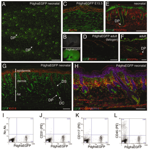
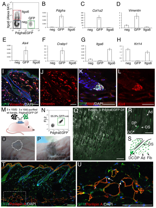
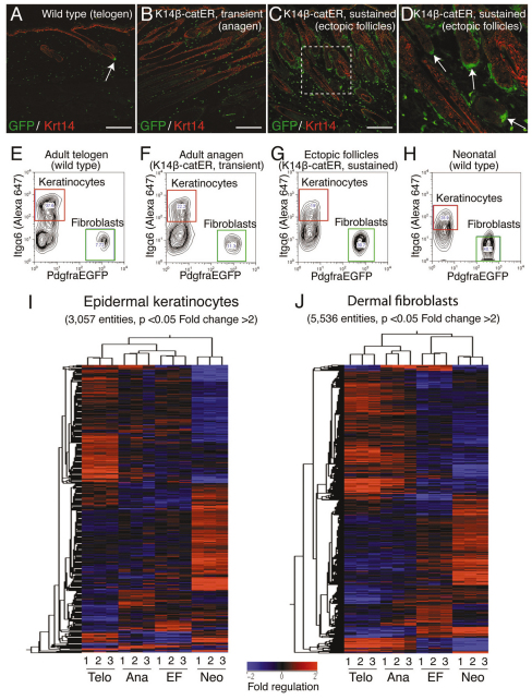
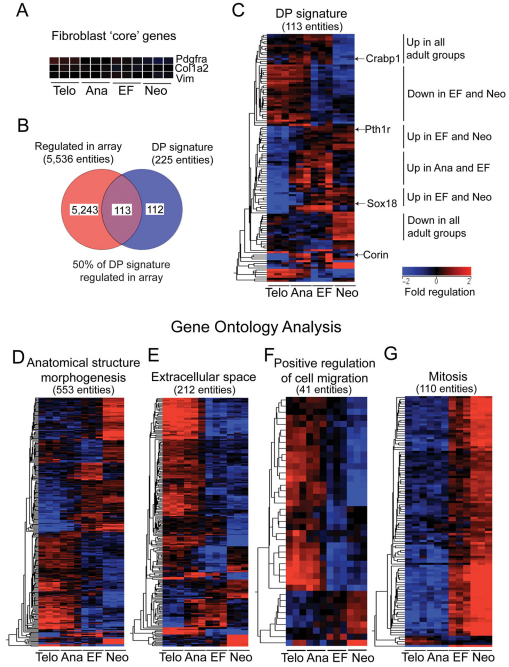
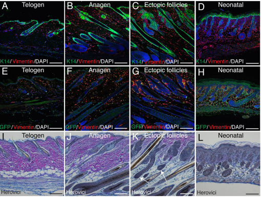
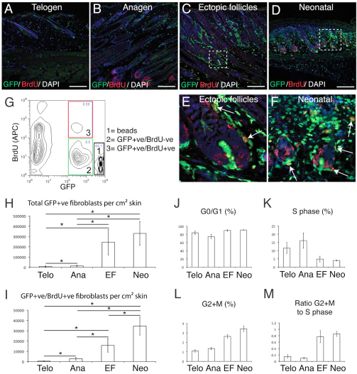
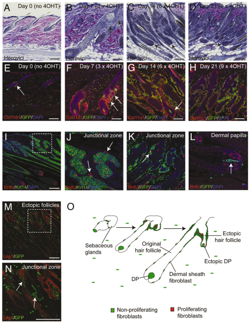
Hair follicle formation depends on reciprocal epidermal-dermal interactions and occurs during skin development, but not in adult life. This suggests that the properties of dermal fibroblasts change during postnatal development. To examine this, we used a PdgfraEGFP mouse line to isolate GFP-positive fibroblasts from neonatal skin, adult telogen and anagen skin and adult skin in which ectopic hair follicles had been induced by transgenic epidermal activation of β-catenin (EF skin). We also isolated epidermal cells from each mouse. The gene expression profile of EF epidermis was most similar to that of anagen epidermis, consistent with activation of β-catenin signalling. By contrast, adult dermis with ectopic hair follicles more closely resembled neonatal dermis than adult telogen or anagen dermis. In particular, genes associated with mitosis were upregulated and extracellular matrix-associated genes were downregulated in neonatal and EF fibroblasts. We confirmed that sustained epidermal β-catenin activation stimulated fibroblasts to proliferate to reach the high cell density of neonatal skin. In addition, the extracellular matrix was comprehensively remodelled, with mature collagen being replaced by collagen subtypes normally present only in developing skin. The changes in proliferation and extracellular matrix composition originated from a specific subpopulation of fibroblasts located beneath the sebaceous gland. Our results show that adult dermis is an unexpectedly plastic tissue that can be reprogrammed to acquire the molecular, cellular and structural characteristics of neonatal dermis in response to cues from the overlying epidermis.


Figures

Fig. 1.
Fig. 1.
Expression of PdgfraEGFP in developing and adult mouse skin. (A) Wholemount of neonatal back skin showing GFP fluorescence in dermal fibroblasts and dermal papilla. (B) Fluorescence images of neonatal PdgfraEGFP mouse (bottom) and wild-type littermate (top). (C-E,G) Paraffin sections of PdgfraEGFP back skin labelled with antibodies to GFP (green, C,D,G), Krt14 (red, C,D,G; green, E) or Pdgfrα (red, E). (F) Cryosection of back skin labelled with Pdgfrα (red) and Krt14 (green) antibodies. (H) Cryosection of back skin labelled with Pdgfrα (red), GFP (green) and Krt14 (blue) antibodies. (I-L) Flow cytometry of PdgfraEGFP adult dermal cells labelled with (I) no antibodies, (J) anti-CD31(PE), (K) anti-CD117(PE), or (L) anti-CD45(PE). DC, dermal cup; DP, dermal papilla; DS, dermal sheath. Scale bars: 150 μm in A; 50 μm in C; 200 μm in D,E,G,H; 40 μm in F.
Fig. 2.
Fig. 2.
PdgfraEGFP expression defines the mouse dermal fibroblast population. (A) Flow cytometry sort gates used to purify Itgα6+ cells (epidermal keratinocytes, red box), GFP+ cells (fibroblasts, green box), and Itgα6 GFP cells (black box) from PdgfraEGFP adult telogen back skin. (B-H) qPCR of mRNA levels in sorted cell populations. Genes were normalised to Gapdh. Each bar represents the average of replicates from four mice (± s.e.). neg, GFP+, Itgα6+. (I-L) Paraffin sections of PdgfraEGFP back skin labelled with GFP (green) and Vim (red, I,J) or Alx4 (red, K,L) antibodies, and counterstained with DAPI (blue) to label nuclei. (M) Outline of grafting experiment. wt kerats, wild-type keratinocytes; DF, dermal fibroblasts. (N) Sort gates used to purify GFP+ cells (green box) from PdgfraEGFP neonatal dermis. (O,P) Hair growth at graft sites 30 days post-grafting. The graft site is outlined in O. (Q,R) Fluorescence/brightfield images of graft sites viewed from the dermal side. Note the refractile, lipid-filled adipocytes in Q. (S) The contribution of PdgfraEGFP+ cells to the different dermal compartments. DP, dermal papilla; DC, dermal cup; DS, dermal sheath; Ad, adipocyte; Fib, fibroblast. (T,U) Paraffin section of PdgfraEGFP back skin labelled with GFP (green) and perilipin A (red) antibodies, and counterstained with DAPI (blue) to label nuclei. The boxed region in T is enlarged in U. Note that perilipin A+ adipocytes have GFP+ nuclei (arrows). Scale bars: 50 μm in I-L; 150 μm in Q; 500 μm in R; 100 μm in T,U.
Fig. 3.
Fig. 3.
Gene expression profiling of epidermal keratinocytes and dermal fibroblasts from PdgfraEGFP mice. (A-D) Paraffin sections of back skin labelled with GFP (green) and Krt14 (red) antibodies. The boxed region in C is enlarged in D. Arrows point to PdgfraEGFP+ fibroblasts associated with the DP of an original hair follicle (A) and ectopic follicles (D). Scale bars: 200 μm. (E-H) Flow cytometry sort gates used to isolate populations of Itgα6+ keratinocytes (red box) and PdgfraEGFP+ dermal fibroblasts (green box) for microarray analysis. (I,J) Heatmaps representing the hierarchical clustering (based on both entities and samples) of entities that are differentially regulated (P<0.05, fold change greater than 2) between at least one of six possible pairs of groups, in (I) epidermal keratinocytes and (J) dermal fibroblasts. Some genes are represented by multiple entities. Telo, ‘Telogen’ group (wild type); Ana, ‘Anagen’ group (K14β-catER, transient activation); EF, ‘Ectopic follicles’ group (K14β-catER, sustained activation); Neo, ‘Neonatal’ group. (I,J) Bar indicates fold regulation (baseline to median of all samples).
Fig. 4.
Fig. 4.
Sustained epidermal activation of β-catenin reprograms adult dermal fibroblasts toward a neonatal state. (A) Heatmap representations of three ‘core’ fibroblast genes expressed at similar levels in all four experimental groups. (B) Venn diagram constructed from two lists comprising (1) all significantly regulated entities (P<0.05, fold change greater than 2) in the dermal fibroblast array and (2) 225 DP signature genes (Rendl et al., 2005). (C) Hierarchical clustering (based on entities) of 113 regulated DP signature genes derived from the region of overlap in B. (D-G) GO analysis of significantly regulated genes in the dermal fibroblast array experiment. Hierarchical clustering was performed using genes grouped under three selected GO terms (from a total of 275 significantly enriched terms, P<0.05), as indicated. Some genes are represented by multiple entities. Scale bar represents fold regulation (baseline to median of all samples).
Fig. 5.
Fig. 5.
Dermal reprogramming induced by sustained epidermal β-catenin activation. (A-H) Paraffin sections of PdgfraEGFP mouse back skin labelled with K14 (green, A-D), GFP (green, E-H) and Vim (red) antibodies, and stained with DAPI (blue) to label nuclei. Part of E is shown at higher magnification in Fig. 2I. (I-L) Paraffin sections stained using Herovici’s method to identify thick, mature collagen fibres (pink) and immature collagen fibrils (blue). Nuclei are stained grey/blue. Arrows in K indicate ectopic follicles. Scale bars: 200 μm.
Fig. 6.
Fig. 6.
Dermal remodelling is associated with an increase in fibroblast proliferation and population density. (A-F) Paraffin sections of mouse back skin labelled with GFP (green) and BrdU (red) antibodies and counterstained with DAPI. E and F are enlarged regions of C and D, respectively. Arrows point to proliferating PdgfraEGFP+ BrdU+ cells. Scale bars: 200 μm. (G) Flow cytometry gates used to quantify cell number relative to counting beads. (H,I) Absolute numbers per cm2 back skin of (H) total PdgfraEGFP+ cells or (I) proliferating PdgfraEGFP+ BrdU+ cells, under different experimental conditions. Asterisks denote a significant difference between two groups (t-test, P<0.05). (J-M) The percentage of PdgfraEGFP+ fibroblasts in (J) G0/G1, (K) S phase and (L) G2+M cell cycle stages; and (M) the ratio of cells in G2+M to cells in S phase. Average ± s.e. of 4-9 replicates.
Fig. 7.
Fig. 7.
Remodelling of adult dermis originates from a population of fibroblasts in the hair follicle junctional zone. (A-L) Sections of K14β-catER mouse back skin treated with 4-OHT for 0, 7, 14 or 21 days as indicated or for 14 days (I-L). (A-D) Paraffin sections stained using Herovici’s method to identify mature (pink fibres) and immature (fine blue fibrils) collagen. (E-H) Frozen sections labelled with anti-Col11α1 (red) and showing GFP fluorescence (green). Nuclei were stained with DAPI (blue). (I-L) Paraffin sections stained for Krt14 (green) and BrdU (red). (M,N) Frozen section of ectopic follicle-forming K14β-catER back skin labelled with anti-Lrig1 (red) and showing GFP fluorescence (green). The boxed regions in I and M are enlarged in J and N, respectively. Arrows point to: (E) a small population of PdgfraEGFP+ fibroblasts associated with the telogen junctional zone; (B,F) regions of recently regenerated collagen at the junctional zone; (C,D,G,N) ectopic follicles; (J,K) BrdU-labelled junctional zone fibroblasts; and (L) the DP of an original hair follicle, which is not labelled with BrdU. (O) The progressive remodelling of adult dermis originating from fibroblasts associated with the hair follicle junctional zone. Scale bars: 200 μm.

References

    1. Baker C. M., Verstuyf A., Jensen K. B., Watt F. M. (2010). Differential sensitivity of epidermal cell subpopulations to beta-catenin-induced ectopic hair follicle formation. Dev. Biol. 343, 40–50 - PMC - PubMed
    1. Bastide P., Darido C., Pannequin J., Kist R., Robine S., Marty-Double C., Bibeau F., Scherer G., Joubert D., Hollande F., et al. (2007). Sox9 regulates cell proliferation and is required for Paneth cell differentiation in the intestinal epithelium. J. Cell Biol. 178, 635–648 - PMC - PubMed
    1. Biernaskie J., Paris M., Morozova O., Fagan M. B., Marra M., Pevny L., Miller F. D. (2009). SKPs derive from hair follicle precursors and exhibit properties of adult dermal stem cells. Cell Stem Cell 5, 610–623 - PMC - PubMed
    1. Chan E. F., Gat U., McNiff J. M., Fuchs E. (1999). A common human skin tumour is caused by activating mutations in β-catenin. Nat. Genet. 21, 410–413 - PubMed
    1. Chi W. Y., Enshell-Seijffers D., Morgan B. A. (2010). De novo production of dermal papilla cells during the anagen phase of the hair cycle. J. Invest. Dermatol. 130, 2664–2666 - PMC - PubMed

Publication types

Substances