. 2012 Jan 15;302(2):C405-11.
doi: 10.1152/ajpcell.00337.2011. Epub 2011 Oct 26.

PDGF enhances store-operated Ca2+ entry by upregulating STIM1/Orai1 via activation of Akt/mTOR in human pulmonary arterial smooth muscle cells

Affiliations

PDGF enhances store-operated Ca2+ entry by upregulating STIM1/Orai1 via activation of Akt/mTOR in human pulmonary arterial smooth muscle cells

Aiko Ogawa et al. Am J Physiol Cell Physiol. .

Abstract

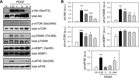
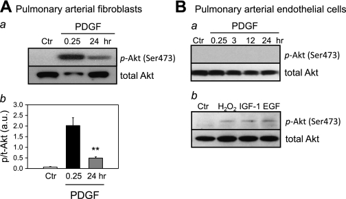
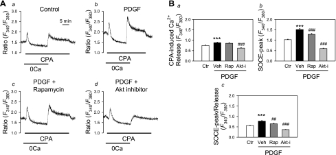
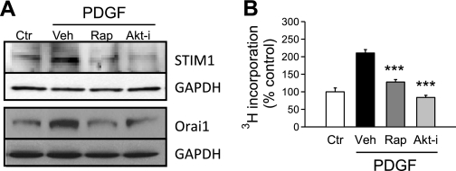
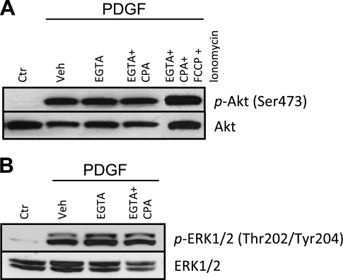
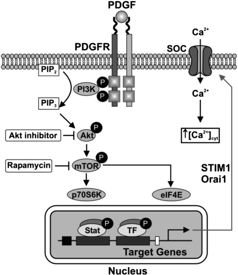
Platelet-derived growth factor (PDGF) and its receptor are known to be substantially elevated in lung tissues and pulmonary arterial smooth muscle cells (PASMC) isolated from patients and animals with pulmonary arterial hypertension. PDGF has been shown to phosphorylate and activate Akt and mammalian target of rapamycin (mTOR) in PASMC. In this study, we investigated the role of PDGF-mediated activation of Akt signaling in the regulation of cytosolic Ca(2+) concentration and cell proliferation. PDGF activated the Akt/mTOR pathway and, subsequently, enhanced store-operated Ca(2+) entry (SOCE) and cell proliferation in human PASMC. Inhibition of Akt attenuated the increase in cytosolic Ca(2+) concentration due to both SOCE and PASMC proliferation. This effect correlated with a significant downregulation of stromal interacting molecule (STIM) and Orai, proposed molecular correlates for SOCE in many cell types. The data from this study present a novel pathway for the regulation of Ca(2+) signaling and PASMC proliferation involving activation of Akt in response to upregulated expression of PDGF. Targeting this pathway may lead to the development of a novel therapeutic option for the treatment of pulmonary arterial hypertension.


Figures

Fig. 1.
Fig. 1.
Platelet-derived growth factor (PDGF)-induced phosphorylation of the Akt/mammalian target of rapamycin (mTOR) pathway in normal human pulmonary arterial smooth muscle cells (PASMC). A: representative Western blots showing time-dependent changes in the phosphorylated [p-Akt, p-mTOR, p-p70S6K (p70 ribosomal S6 kinase), p-4EBP1 (eukaryotic initiation factor 4E binding protein 1), and p-eIF4E (eukaryotic initiation factor 4E)] components and total proteins (t-Akt, t-mTOR, t-p70S6K, t-4EBP1, and t-eIF4E) of the Akt/mTOR signaling pathway induced by PDGF stimulation (10 ng/ml). Protein expression was assessed at 0.25, 3, 12, and 24 h after PDGF treatment. B: summary data (means ± SE, n = 3) were quantitated and compared at each time point, with the control (Ctr) being without PDGF treatment. au, Arbitrary units. *P < 0.05, **P < 0.01, and ***P < 0.001 vs. Ctr.
Fig. 2.
Fig. 2.
Akt phosphorylation in pulmonary arterial fibroblasts (PAF) and pulmonary arterial endothelial cells (PAEC). A, a: representative Western blots showing time-dependent changes in the p-Akt and t-Akt induced by PDGF stimulation (10 ng/ml). Protein expression was assessed at 0.25 and 24 h after PDGF treatment. b: Summarized data (means ± SE, n = 3) were quantitated and compared at each time point. **P < 0.01 vs. Ctr. B, a: representative Western blots showing no phosphorylation of Akt induced by PDGF stimulation (10 ng/ml). b: Treatment with hydrogen peroxide (500 μM), insulin-like growth factor-I (IGF-I; 50 ng/ml), and EGF (50 ng/ml) for 0.25 h induced phosphorylation of Akt. Blots were representative of 3 different sets of experiments.
Fig. 3.
Fig. 3.
Inhibition of Akt/mTOR attenuates PDGF-induced increase in store-operated Ca2+ entry (SOCE) in PASMC. A: representative traces showing changes in cytosolic Ca2+ concentration ([Ca2+]cyt) [expressed as the ratio of the 340 and 380 fluorescence (F340/F380)] in response to cyclopiazonic acid (CPA)-induced store depletion in the absence of extracellular Ca2+ and subsequent SOCE upon replenishment of extracellular Ca2+. Cells were treated with PDGF and other inhibitors [a: Ctr; b: PDGF (10 ng/ml), c: PDGF and rapamycin (Rap; 10 nM), d: PDGF and Akt inhibitor (Akt-I; 1 μM)] for 24 h before Ca2+ measurement. B: quantitative comparison of the CPA-induced Ca2+ release (a), the peak of SOCE (SOCE-peak; b), and the ratio of SOCE-peak to CPA-induced Ca2+ release (SOCE-peak/release; c) in Ctr (n = 65), PDGF + vehicle (Veh, n = 64), PDGF + Rap (n = 50), and PDGF + Akt-i (n = 58) PASMC. Rap and Akt-i both significantly attenuated PDGF-induced augmentation of SOCE. Values are means ± SE. ***P < 0.001 vs. Ctrl. ##P < 0.01, ###P < 0.001 vs. PDGF + Veh-treated cells.
Fig. 4.
Fig. 4.
Inhibition of Akt/mTOR blocks PDGF-induced stromal interacting molecule 1 (STIM1)/Orai1 expression and PASMC proliferation. A: representative blots showing PDGF treatment (24 h)-induced upregulation of STIM1 and Orai1 expression in normal PASMC. After treatment with Rap (10 nM) or Akt-i (1 μM), PDGF-induced expression of both molecules was decreased. Experiments were repeated 3 times. B: bar charts comparing the effect of Rap (10 nM) and Akt-i (1 μM) on the PDGF-stimulated proliferation of normal PASMC using 3H incorporation (in counts per minute). ***P < 0.001 vs. PDGF + Veh-treated cells.
Fig. 5.
Fig. 5.
Phosphorylation of Akt induced by PDGF is independent of extracellular and intracellular Ca2+. PASMC were treated for 0.25 h with PDGF (10 ng/ml) and/or Veh, EGTA (2 mM), CPA (10 μM), carbonyl cyanide-p-trifluoromethoxyphenylhydrazone (FCCP; 5 μM), or ionomycin (10 μM) to chelate Ca2+. A: representative Western blots showing the expression level of p- and t-Akt. Blots were representative of 3 different sets of experiments. B: representative Western blots showing the expression level of p- and t-ERK1/2. Blots were representative of 3 different sets of experiments.
Fig. 6.
Fig. 6.
Schematic of proposed pathway. Binding of PDGF to its receptor (PDGFR) activates Akt/mTOR signaling, leading to increased expression of STIM1/Orai1, increased SOCE, and increased cell proliferation. SOC, store-operated Ca2+ channels; PIP2, phosphatidylinositol 4,5-bisphosphate; PIP3, phosphatidylinositol (3,4,5)-trisphosphate; TF, transcription factor.

References

    1. Balciunaite E, Jones S, Toker A, Kazlauskas A. PDGF initiates two distinct phases of protein kinase C activity that make unequal contributions to the G0 to S transition. Curr Biol 10: 261–267, 2000 - PubMed
    1. Barst RJ. PDGF signaling in pulmonary arterial hypertension. J Clin Invest 115: 2691–2694, 2005 - PMC - PubMed
    1. Bisaillon JM, Motiani RK, Gonzalez-Cobos JC, Potier M, Halligan KE, Alzawahra WF, Barroso M, Singer HA, Jourd'heuil D, Trebak M. Essential role for STIM1/Orai1-mediated calcium influx in PDGF-induced smooth muscle migration. Am J Physiol Cell Physiol 298: C993–C1005, 2010 - PMC - PubMed
    1. Bonnet S, Paulin R, Sutendra G, Dromparis P, Roy M, Watson KO, Nagendran J, Haromy A, Dyck JRB, Michelakis ED. Dehydroepiandrosterone reverses systemic vascular remodeling through the inhibition of the Akt/GSK3-β/NFAT axis. Circulation 120: 1231–1240, 2009 - PubMed
    1. Cahalan MD. STIMulating store-operated Ca(2+) entry. Nat Cell Biol 11: 669–677, 2009 - PMC - PubMed

Publication types

MeSH terms

LinkOut - more resources