. 2011 Nov 8;108(45):18536-41.
doi: 10.1073/pnas.1111597108. Epub 2011 Oct 26.

Psb27, a transiently associated protein, binds to the chlorophyll binding protein CP43 in photosystem II assembly intermediates

Affiliations

Psb27, a transiently associated protein, binds to the chlorophyll binding protein CP43 in photosystem II assembly intermediates

Haijun Liu et al. Proc Natl Acad Sci U S A. .

Abstract

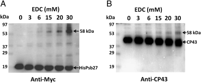
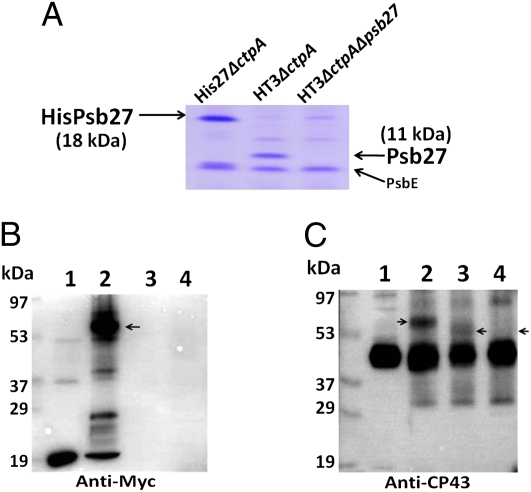
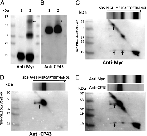
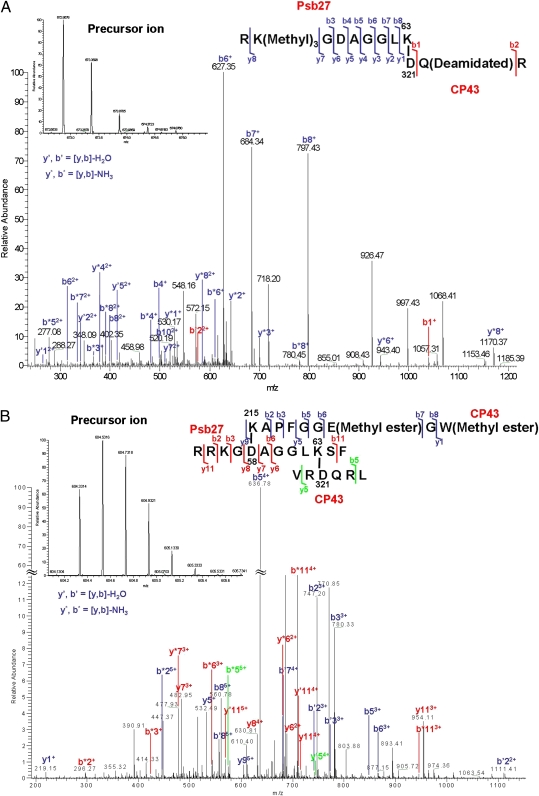
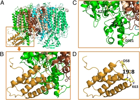
Photosystem II (PSII), a large multisubunit pigment-protein complex localized in the thylakoid membrane of cyanobacteria and chloroplasts, mediates light-driven evolution of oxygen from water. Recently, a high-resolution X-ray structure of the mature PSII complex has become available. Two PSII polypeptides, D1 and CP43, provide many of the ligands to an inorganic Mn(4)Ca center that is essential for water oxidation. Because of its unusual redox chemistry, PSII often undergoes degradation followed by stepwise assembly. Psb27, a small luminal polypeptide, functions as an important accessory factor in this elaborate assembly pathway. However, the structural location of Psb27 within PSII assembly intermediates has remained elusive. Here we report that Psb27 binds to CP43 in such assembly intermediates. We treated purified genetically tagged PSII assembly intermediate complexes from the cyanobacterium Synechocystis 6803 with chemical cross-linkers to examine intermolecular interactions between Psb27 and various PSII proteins. First, the water-soluble 1-ethyl-3-(3-dimethylaminopropyl)carbodiimide (EDC) was used to cross-link proteins with complementary charged groups in close association to one another. In the His27△ctpAPSII preparation, a 58-kDa cross-linked species containing Psb27 and CP43 was identified. This species was not formed in the HT3△ctpA△psb27PSII complex in which Psb27 was absent. Second, the homobifunctional thiol-cleavable cross-linker 3,3'-dithiobis(sulfosuccinimidylpropionate) (DTSSP) was used to reversibly cross-link Psb27 to CP43 in His27△ctpAPSII preparations, which allowed the use of liquid chromatography/tandem MS to map the cross-linking sites as Psb27K(63)↔CP43D(321) (trypsin) and CP43K(215)↔Psb27D(58)AGGLK(63)↔CP43D(321) (chymotrypsin), respectively. Our data suggest that Psb27 acts as an important regulatory protein during PSII assembly through specific interactions with the luminal domain of CP43.


Conflict of interest statement

The authors declare no conflict of interest.

Figures

Fig. 1.
Fig. 1.
EDC cross-linking of Psb27 and CP43. Concentration-dependent production of cross-linked species at 58 kDa is shown. His27△ctpAPSII complex was treated with EDC at various concentrations, and immunoblots were probed with anti-Myc antibodies that recognize the Myc epitope on the tagged Psb27 protein (A) or anti-CP43 antibodies (B). EDC concentrations are shown at the top of each blot. The locations of molecular mass standards are shown on the left. Cross-linked products and the parent polypeptides are identified on the right.
Fig. 2.
Fig. 2.
EDC cross-linking in three types of purified PSII complexes. (A) SDS/PAGE analysis showing slower migration of His/Myc-tagged Psb27 (18 kDa, lane 1) than native Psb27 (11 kDa, lane 2) after Coomassie Brilliant Blue (R-250) staining. Lane 1: His27△ctpAPSII; lane 2: HT3△ctpAPSII; lane 3: HT3△ctpApsb27PSII. Two micrograms of chlorophyll a-containing samples were loaded in each lane. (B) Detection of cross-linked species. Lane 1: His27△ctpAPSII; lane 2: His27△ctpAPSII + EDC; lane 3: HT3△ctpAPSII + EDC; lane 4: HT3△ctpApsb27PSII + EDC. EDC (30 mM) cross-linking reaction was performed for 30 min in the dark at 23 °C. Immunoblot was probed with anti-Myc antibodies. (C) Same as in B except probed with anti-CP43 antibodies. Arrows indicate the increased mobility of CP43–Psb27 cross-linked complex (lane 3) compared with His/Myc-tagged Psb27–CP43 cross-linked complex (lane 2) and the absence of any cross-linked product in lane 4 because of the absence of Psb27 in HT3△ctpApsb27PSII.
Fig. 3.
Fig. 3.
DTSSP cross-linking in His27△ctpAPSII preparation. (A) Lane 1: His27△ctpAPSII; lane 2: His27△ctpAPSII + DTSSP. The cross-linking reaction was performed by using 10 mM DTSSP for 30 min in the dark at 23 °C. Immunoblot was probed with anti-Myc antibodies. The location of the 58-kDa CP43–Psb27 cross-linked species is indicated with an arrow. (B) Same as in A except probed with anti-CP43 antibodies. (C) Results of 2D diagonal electrophoresis of His27△ctpAPSII complex treated with DTSSP and probed with anti-Myc antibodies. During SDS/PAGE, β-mercaptoethanol was omitted in the first dimension and was included in the second dimension. (D) Same as in C except probed with anti-CP43 antibodies. (E) Overlay of C and D. Protein spots that represent tagged Psb27 and CP43 are found as a vertical series below the 58-kDa cross-linking complex, indicating intermolecular cross-links between these two components.
Fig. 4.
Fig. 4.
Product ion (tandem MS) spectra obtained for cross-linked peptides. (A) Product ion (tandem MS) spectra of the cross-linked peptides RKGDAGGLK63 (Psb27) and D321QR (CP43). The fragmentation pattern indicates that cross-links are formed specifically between K63 of Psb27 peptide 55–63 and D321 of CP43 peptide 321–323. Shown is the product ion map of cross-link fragment after collision-induced dissociation. The abundant b′, y′ and b*, y* ions are labeled. Ions showing cross-linking were m/z = 672.8678 (2+) (Inset). (B) Product ion (tandem MS) spectra of the intermolecular cross-links after chymotrypsin digestion. Peptide 54–65 of Psb27 protein was sandwich–cross-linked to peptide 215–223 and 319–324 of CP43, respectively. Cross-links were formed specifically between K215 of CP43 peptide 215–223 and D58 of Psb27 peptide 54–65 and between K63 of Psb27 peptide 54–65 and D321 of CP43 peptide 319–324, respectively. Ions showing cross-linking were of m/z = 604.3314 (5+) (Inset). The abundant b′, y′ and b*, y* ions are labeled.
Fig. 5.
Fig. 5.
A schematic model of binding of Psb27 to the luminal domain of CP43 (PDB IDs 3ARC and 2KMF). (A) Front view showing Psb27 (bright orange) binding to loop C and loop E of CP43 (green). D1, brown; D2, cyan; CP47, light green; small subunit peptides, wheat. (B) A magnified view of the model shown in A. CP43K215, Psb27D58, Psb27K63, and CP43D321 are represented as sticks. (C) Distance between K215 and D321 of CP43 in the X-ray structural model (PDB ID 3ARC). (D) Distance between D58 and K63 of Psb27 in the NMR model (PDB ID 2KMF, model 1). All images were prepared with PyMOL software (30).

References

    1. Umena Y, Kawakami K, Shen JR, Kamiya N. Crystal structure of oxygen-evolving photosystem II at a resolution of 1.9 Å. Nature. 2011;473:55–60. - PubMed
    1. Nowaczyk MM, et al. Psb27, a cyanobacterial lipoprotein, is involved in the repair cycle of photosystem II. Plant Cell. 2006;18:3121–3131. - PMC - PubMed
    1. Roose JL, Pakrasi HB. The Psb27 protein facilitates manganese cluster assembly in photosystem II. J Biol Chem. 2008;283:4044–4050. - PubMed
    1. Ikeuchi M, Inoue Y, Vermaas W. Characterization of photosystem II subunits from the cyanobacterium Synechocystis sp. PCC6803. In: Mathis P, editor. Photosynthesis: From Light to Biosphere. Vol III. Dordrecht, The Netherlands: Kluwer; 1995. pp. 297–300.
    1. Kashino Y, et al. Proteomic analysis of a highly active photosystem II preparation from the cyanobacterium Synechocystis sp. PCC 6803 reveals the presence of novel polypeptides. Biochemistry. 2002;41:8004–8012. - PubMed

Publication types

MeSH terms

Substances

LinkOut - more resources