. 2011;6(10):e24899.
doi: 10.1371/journal.pone.0024899. Epub 2011 Oct 21.

Automated detection and segmentation of synaptic contacts in nearly isotropic serial electron microscopy images

Affiliations

Automated detection and segmentation of synaptic contacts in nearly isotropic serial electron microscopy images

Anna Kreshuk et al. PLoS One. 2011.

Abstract

We describe a protocol for fully automated detection and segmentation of asymmetric, presumed excitatory, synapses in serial electron microscopy images of the adult mammalian cerebral cortex, taken with the focused ion beam, scanning electron microscope (FIB/SEM). The procedure is based on interactive machine learning and only requires a few labeled synapses for training. The statistical learning is performed on geometrical features of 3D neighborhoods of each voxel and can fully exploit the high z-resolution of the data. On a quantitative validation dataset of 111 synapses in 409 images of 1948×1342 pixels with manual annotations by three independent experts the error rate of the algorithm was found to be comparable to that of the experts (0.92 recall at 0.89 precision). Our software offers a convenient interface for labeling the training data and the possibility to visualize and proofread the results in 3D. The source code, the test dataset and the ground truth annotation are freely available on the website http://www.ilastik.org/synapse-detection.


Conflict of interest statement

Competing Interests: The authors have read the journal's policy and have the following conflicts. This study was partially funded by Robert Bosch GmbH. There are no patents, products in development or marketed products to declare. This does not alter the authors' adherence to all the PLoS ONE policies on sharing data and materials, as detailed online in the guide for authors.

Figures

Figure 1
Figure 1. User labels and algorithm predictions.
Top row: the complete set of user annotations for the first training set (20 brush strokes in total), with yellow labels for synapses, red for membranes, green for the rest. Bottom row: raw data and algorithm predictions on two other slices in the first training set. In black circles: some unlabeled synapses and their probability maps. The color intensity corresponds to the certainty in the prediction, predictions for green class are omitted for clarity.
Figure 2
Figure 2. Precision and recall of the algorithm and the human experts.
Recall was calculated as the (no. of true positives)/(no. of synapses in the ground truth), precision as the (no. of true positives)/(total no. of synapse candidates). A: Precision and recall of the algorithm results for the four different training sets. B: Precision and recall of the algorithm compared to the human experts with and without the time limit. The synapse probability threshold values are annotated next to the corresponding points of the curve.
Figure 3
Figure 3. 3D visualization of the results.
Top: all synapses detected by the algorithm after training on the labels from Fig. 1. Bottom: a close-up view of three differently oriented synapses.
Figure 4
Figure 4. Synapse detection summary report.
Part of the summary report produced by ilastik. The fourth detection from the top (no. 36) is a false positive, which can easily be filtered out by a human expert by looking at a larger context.
Figure 5
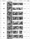
Figure 5. Error examples.
A, B, C: false negative decisions of the human observers, D, E, F: false positive detections of the human observers, shown as yellow “ball” labels in the image center, G, H, I: false negative decisions of the algorithm, J, K, L false positive decisions of the algorithm.

References

    1. Sterio DC. The unbiased estimation of number and sizes of arbitrary particles using the disector. Journal of Microscopy. 1984;134:127–136. - PubMed
    1. Geinisman Y, Gundersen HJG, Zee E, West MJ. Unbiased stereological estimation of the total number of synapses in a brain region. Journal of Neurocytology. 1996;25:805–819. - PubMed
    1. Mayhew TM. How to count synapses unbiasedly and efficiently at the ultrastructural level: proposal for a standard sampling and counting protocol. Journal of Neurocytology. 1996;25:793–804. - PubMed
    1. Coggeshall RE, Lekan HA. Methods for determining numbers of cells and synapses: A case for more uniform standards of review. The Journal of Comparative Neurology. 1996;364:6–15. - PubMed
    1. Kubota Y. Important factors for the three-dimensional reconstruction of neuronal structures from serial ultrathin sections. Frontiers in Neural Circuits. 2009;3 - PMC - PubMed

Publication types