Review
. 2012 Jan 15;302(2):F227-33.
doi: 10.1152/ajprenal.00561.2011. Epub 2011 Oct 26.

The first decade of using multiphoton microscopy for high-power kidney imaging

Affiliations
Review

The first decade of using multiphoton microscopy for high-power kidney imaging

János Peti-Peterdi et al. Am J Physiol Renal Physiol. .

Abstract

In this review, we highlight the major scientific breakthroughs in kidney research achieved using multiphoton microscopy (MPM) and summarize the milestones in the technological development of kidney MPM during the past 10 years. Since more and more renal laboratories invest in MPM worldwide, we discuss future directions and provide practical, useful tips and examples for the application of this still-emerging optical sectioning technology. Advantages of using MPM in various kidney preparations that range from freshly dissected individual glomeruli or the whole kidney in vitro to MPM of the intact mouse and rat kidney in vivo are reviewed. Potential combinations of MPM with micromanipulation techniques including microperfusion and micropuncture are also included. However, we emphasize the most advanced and complex, quantitative in vivo imaging applications as the ultimate use of MPM since the true mandate of this technology is to look inside intact organs in live animals and humans.


Figures

Fig. 1.
Fig. 1.
Timeline of the development of multiphoton microscopy (MPM) including its use for kidney research. The first practical demonstration of MPM (5) and the development of single-box femtosecond Ti:Sapphire lasers (4) took place about 20 years ago. The first commercial MPM system was built by Bio-Rad a few years later, shortly after enhanced green fluorescent protein (GFP) became available (12). The first applications of MPM in the kidney came about the same time as in other organs with the publication of its use on fixed kidney (42), ex vivo (37, 38), and in vivo (6) about 10 years ago. Superresolution microscopy, for example, stimulated emission depletion (STED) became available more recently, further pushing the limits of fluorescence microscopy.
Fig. 2.
Fig. 2.
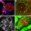
MPM imaging of hardly accessible kidney cell types and structures. Multicolor imaging of the juxtaglomerular apparatus (JGA) in vitro (A; adapted from Ref. 32) using the freshly dissected and microperfused rabbit glomerulus (G) with attached afferent arteriole (AA) and macula densa (MD), and in vivo (B) in the intact Munich-Wistar-Fromter rat kidney. Individual renin granules in the final part of the AA were labeled green using quinacrine. In addition, the cell membrane marker R-18 (red) and nuclear label Hoechst 33342 (blue) were used (in A). The plasma was labeled with 70-kDa dextran-rhodamine B (red) visible within the G, AA, and efferent arteriole (EA; in B). Using the same techniques, podocytes (arrows) can be directly visualized in the microperfused glomerulus in vitro (C; adapted from Ref. 47) or in vivo (D). In a Munich-Wistar-Fromter rat (D), 70-kDa dextran-rhodamine B labeled the plasma (red), and Lucifer yellow, a 0.4-kDa easily filterable, but cell membrane impermeable small molecule (green) was infused continuously into the carotid artery to label the primary filtrate in Bowman's space and the proximal tubule (PT) green. Since podocytes (arrows) and parietal cells normally do not endocytose Lucifer yellow, they remain unlabeled (dark, negative image). Scale bars = 20 μm if not specified.
Fig. 3.
Fig. 3.
MPM of cytosolic parameters in the kidney in vivo. Micropuncture delivery of Rhod 2-AM (A) or SNARF-1-AM (B) directly into the tubular fluid in different nephron segments in Munich-Wistar-Fromter rats allowed direct visualization of intracellular calcium (A) or pH (B; both are red), respectively. Plasma was labeled green using 500-kDa dextran-FITC. A: 2 adjacent nephron's connecting tubule segments (CNT) merge into a common cortical collecting duct (CCD). B: intense SNARF-1 fluorescence is observed in select bulging epithelial cells of the CCD (arrows, most likely type A intercalated cells). PT, proximal tubule. Scale bar = 20 μm.
Fig. 4.
Fig. 4.
MPM imaging of glomerular pathology in vivo. In a puromycin aminonucleoside (PAN)-induced model of focal segmental glomerulosclerosis (FSGS) in Munich-Wistar-Fromter rats, the intravascular space (plasma) marker Alexa 594-albumin (red) was given in a bolus, and Lucifer yellow was infused continuously into the carotid artery to label the primary filtrate in Bowman's space (yellowish green). Numerous large cysts in dark, unlabeled podocytes (asterisk) are visible after PAN treatment (A). The nuclear stain Hoechst 33342 was given (iv) to help the identification of various cells (B). Examination of the linear profile of Alexa 594-labeled albumin fluorescence intensity in Bowman's space around glomerular capillaries (indicated by white line in B) suggests that the increased albumin permeability of the glomerular filtration barrier in this model is restricted to focal areas (C). Scale bar = 20 μm.
Fig. 5.
Fig. 5.
MPM imaging of cell lineage in vivo in transgenic mouse models. In Tie-2 GFP mice, which express the enhanced GFP in the vascular endothelium, the peritubular capillaries in the renal cortex are visualized in green. The intravascular space (plasma) was labeled with Alexa 594-albumin (red). Scale bar = 20 μm.
Fig. 6.
Fig. 6.
Example for the use of the MP laser as a micromanipulator tool. In a Munich-Wistar-Fromter rat, the intravascular space (plasma) was labeled with Alexa 594-albumin (red). After a control image (A) was taken, a high-power laser beam was focused on one small part of a glomerular capillary (labeled X), which caused disruption of the capillary wall and single-nephron hematuria (B). The plasma and the unlabeled red blood cells are seen entering Bowman's space and PT segments. The distal convoluted tubule (DCT) was labeled green by a previous Lucifer yellow bolus injection into the carotid artery.

References

    1. Basile DP, Friedrich JL, Spahic J, Knipe N, Mang H, Leonard EC, Changizi-Ashtiyani S, Bacallao RL, Molitoris BA, Sutton TA. Impaired endothelial proliferation and mesenchymal transition contribute to vascular rarefaction following acute kidney injury. Am J Physiol Renal Physiol 300:F721–F733, 2011 - PMC - PubMed
    1. Bell PD, Lapointe JY, Sabirov R, Hayashi S, Peti-Peterdi J, Manabe KI, Kovacs G, Okada Y. Macula densa cell signaling involves ATP release through a maxi anion channel. Proc Natl Acad Sci USA 100:4322–4327, 2003 - PMC - PubMed
    1. Comper WD, Osicka TM, Russo LM. Renal filtration, transport, and metabolism of albumin and albuminuria. In: Seldin and Giebisch's The Kidney Physiology and Pathophysiology, edited by Alpern RJ, Hebert SC. New York: Academic, 2008, p. 2081–2112
    1. Curley PF, Ferguson AI. Actively mode-locked Ti:sapphire laser producing transform-limited pulses of 150-fs duration. Opt Lett 16:1016–1018, 1991 - PubMed
    1. Denk W, Strickler J, Webb W. Two-photon laser scanning fluorescence microscopy. Science 248:73–76, 1990 - PubMed

Publication types

MeSH terms

LinkOut - more resources