. 2011 Oct 26;31(43):15376-83.
doi: 10.1523/JNEUROSCI.2271-11.2011.

State-dependent and timing-dependent bidirectional associative plasticity in the human SMA-M1 network

Affiliations

State-dependent and timing-dependent bidirectional associative plasticity in the human SMA-M1 network

Noritoshi Arai et al. J Neurosci. .

Abstract

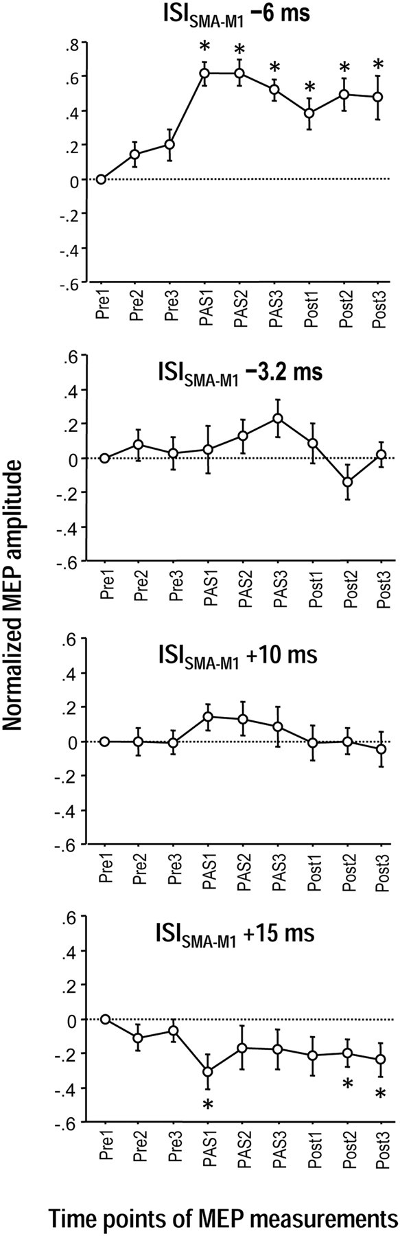
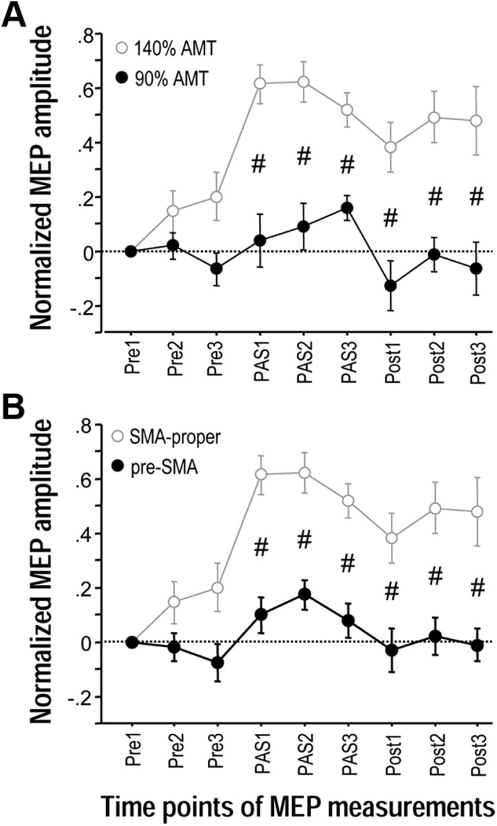
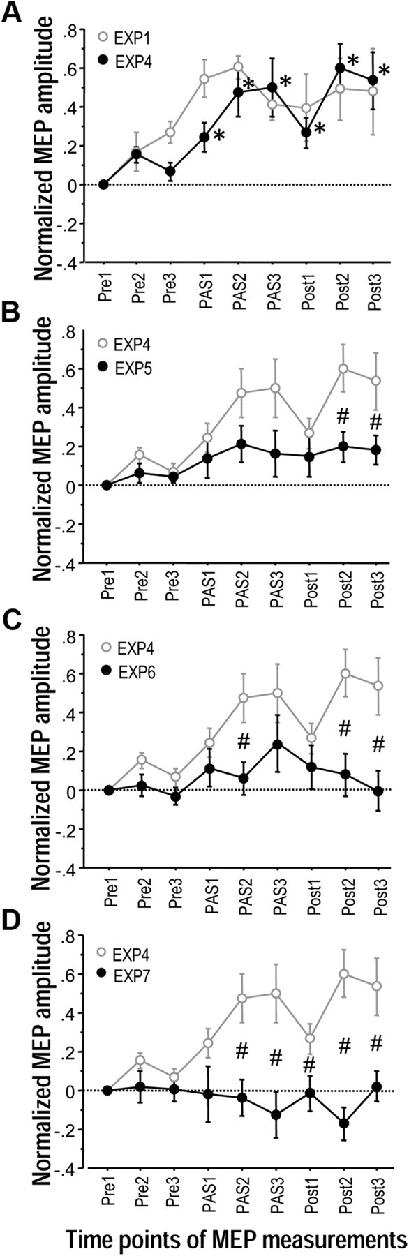
The supplementary motor area (SMA-proper) plays a key role in the preparation and execution of voluntary movements. Anatomically, SMA-proper is densely reciprocally connected to primary motor cortex (M1), but neuronal coordination within the SMA-M1 network and its modification by external perturbation are not well understood. Here we modulated the SMA-M1 network using MR-navigated multicoil associative transcranial magnetic stimulation in healthy subjects. Changes in corticospinal excitability were assessed by recording motor evoked potential (MEP) amplitude bilaterally in a hand muscle. We found timing-dependent bidirectional Hebbian-like MEP changes during and for at least 30 min after paired associative SMA-M1 stimulation. MEP amplitude increased if SMA stimulation preceded M1 stimulation by 6 ms, but decreased if SMA stimulation lagged M1 stimulation by 15 ms. This associative plasticity in the SMA-M1 network was highly topographically specific because paired associative stimulation of pre-SMA and M1 did not result in any significant MEP change. Furthermore, associative plasticity in the SMA-M1 network was strongly state-dependent because it required priming by near-simultaneous M1 stimulation to occur. We conclude that timing-dependent bidirectional associative plasticity is demonstrated for the first time at the systems level of a human corticocortical neuronal network. The properties of this form of plasticity are fully compatible with spike-timing-dependent plasticity as defined at the cellular level. The necessity of priming may reflect the strong interhemispheric connectivity of the SMA-M1 network. Findings are relevant for better understanding reorganization and potentially therapeutic modification of neuronal coordination in the SMA-M1 network after cerebral lesions such as stroke.


Figures

Figure 1.
Figure 1.
Methodology and experimental design. A, Multicoil focal TMS. Near-simultaneous bilateral M1 stimulation (ISI = 0.8 ms; left M1 always before right M1, stimulus intensities adjusted to elicit MEP of 1 mV peak-to-peak amplitude in the contralateral FDI) was delivered through 50 mm diameter figure-of-eight coils. During paired associative stimulation (PAS1–3, see C), SMA-proper was stimulated time-locked to bilateral M1 stimulation using a small 25 mm figure-of-eight coil (stimulus intensity set to 140% of AMT as determined over left M1). Directions of induced currents in the brain were as indicated by the white arrows. For further details, see Materials and Methods. B, Sagittal view at x = −4 in Talairach stereotaxic space (Talairach and Tournoux, 1988) of the mean anatomical image indicating SMA-proper stimulation sites (center of stimulating coil, red circles) and pre-SMA stimulation sites (yellow circles) during PAS1–3 in those subjects (n = 7 for SMA-proper, n = 4 for pre-SMA) in whom MR-navigation was used to verify stimulation topography. Circles are artificially stacked along the z-axis to provide visibility of all individual stimulation sites. The vertical plane through the anterior commissure (white line, y = 0) and the perpendicular anterior–posterior commissure plane (black line) are indicated. Mean coordinates for the SMA-proper stimulation sites were y = −1.3 ± 0.6 (Talairach and Tournoux, 1988) and 3.3 ± 0.2 cm anterior to Cz, mean coordinates for the pre-SMA stimulation sites were y = 23.7 ± 3.3 (Talairach and Tournoux, 1988) and 6.2 ± 3.2 cm anterior to Cz. C, General time line of Experiment 1–7, always consisting of nine blocks of 50 trials each (Pre1–3, PAS1–3, Post1–3). Pre1–3 measure M1 excitability at baseline, PAS1–3 apply paired associative stimulation of SMA and M1, and Post1–3 assess any lasting effects on M1 excitability after PAS1–3. Variations of the stimulation procedures in Experiments 1–7 are detailed in Materials and Methods. EXP, Experiment; Bi-M1, bilateral M1 stimulation; L-M1, left M1 stimulation; SMA, SMA-proper.
Figure 2.
Figure 2.
Timing-dependent associative plasticity between SMA and bilateral M1 (Experiments 1). MEP changes before (Pre1–3), during (PAS1–3) and after (Post1–3) associative stimulation of SMA paired with near-simultaneous stimulation of bilateral M1. MEP amplitudes increased significantly during and for at least 30 min after PAS1–3 at the ISI of −6 ms (SMA before bilateral M1 stimulation, top). In contrast, MEP amplitudes decreased for at least 30 min after PAS1–3 at the ISI of +15 ms (SMA after bilateral M1 stimulation, bottom panel). No significant MEP changes occurred with the ISI of −3.2 ms (second panel from top) and the ISI of +10 ms (second panel from bottom). The MEP changes at the ISI of −6 ms were significantly different from all other ISIs, those at the ISI of −3.2 ms were different from the ISI of +15 ms, and those of the ISI of +10 ms were different from the ISI of +15 ms (all p < 0.009). All MEP data were pooled from the left and right M1 because the rmANOVAs did not show an effect of Side (left vs right M1) or its interactions with any other main effect. Asterisks denote significant MEP changes compared with Pre1 (p < 0.05, post hoc t tests). MEP data in this and all following figures are logarithmized and normalized to Pre1, and shown as means ± SEM.
Figure 3.
Figure 3.
Effects of SMA stimulation intensity and topographic specificity of associative plasticity between SMA and bilateral M1 (Experiments 2 and 3). A, MEP changes before (Pre1–3), during (PAS1–3) and after (Post1–3) associative stimulation of SMA paired with near-simultaneous stimulation of bilateral M1. The intensity of SMA stimulation was reduced to 90% of AMT (black circles). Data are compared with 140% of AMT (gray circles, data from Experiment 1). The ISI between SMA and bilateral M1 stimulation was −6 ms. B, MEP changes before (Pre1–3), during (PAS1–3) and after (Post1–3) associative stimulation of SMA paired with near-simultaneous stimulation of bilateral M1 at a stimulation site 3 cm anterior to the individually determined SMA-proper (i.e., corresponding to pre-SMA, black circles). Stimulation intensity was 140% of AMT, and ISI between SMA and bilateral M1 stimulation was −6 ms. Data are compared with SMA-proper stimulation (gray circles, data from Experiment 1). MEP data were pooled from the left and right M1 because the rmANOVAs did not show an effect of Side (left vs right M1) or its interactions with any other main effect. Significant difference of MEP changes at a single time point between the experimental conditions, #p < 0.005, unpaired t tests. Note that lasting MEP increase critically depends on efficient stimulation of SMA-proper and does not occur with low stimulation intensity or stimulation of pre-SMA.
Figure 4.
Figure 4.
Priming by near-synchronous bilateral M1 stimulation is necessary for induction of associative plasticity between SMA and M1 (Experiments 4–7). A, MEP changes in left M1 before (Pre1–3), during (PAS1–3) and after (Post1–3) paired associative stimulation of SMA and unilateral left M1 (Experiment 4, black circles). The ISI between SMA and left M1 stimulation in this and all other experiments of this Figure was −6 ms. During Pre1–3 near-simultaneous bilateral M1 stimulation was conducted, during Post1–3 only left M1 was stimulated. Data are compared with paired associative stimulation of SMA with bilateral M1 (Experiment 1, gray circles). Note that there was no difference in MEP change between experimental conditions. In both conditions, MEP amplitude increased significantly during and for at least 30 min after PAS1–3. Asterisks denote significant MEP changes compared with Pre1 in Experiment 4 (p < 0.05). B, MEP changes in left M1 before (Pre1–3), during (PAS1–3) and after (Post1–3) paired associative stimulation of SMA and unilateral left M1. During Pre1–3 and Post1–3, only the left M1 was stimulated (Experiment 5, black circles). Here and in C and D, MEP data are compared with those from Experiment 4 (gray circles). Here and in C and D, #p < 0.05, significant difference in MEP changes at single time points between the experimental conditions. C, MEP changes in left M1 before (Pre1–3), during (PAS1–3) and after (Post1–3) associative stimulation of SMA paired with near-simultaneous stimulation of bilateral M1. During Pre1–3 and Post1–3, only the left M1 was stimulated (Experiment 6, black circles). D, MEP changes in left M1 before (Pre1–3), during (PAS1–3) and after (Post1–3) paired associative stimulation of SMA and unilateral left M1. During Pre1–3, nonsimultaneous bilateral M1 stimulation was delivered (ISI = 5 ms), during Post1–3, only the left M1 was stimulated (Experiment 7, black circles). Of note, lasting MEP increase occurred only if paired associative stimulation of the SMA-M1 network was primed by near-simultaneous bilateral M1 stimulation during Pre1–3.

References

    1. Bestmann S, Baudewig J, Siebner HR, Rothwell JC, Frahm J. Subthreshold high-frequency TMS of human primary motor cortex modulates interconnected frontal motor areas as detected by interleaved fMRI-TMS. Neuroimage. 2003;20:1685–1696. - PubMed
    1. Bi G, Poo M. Synaptic modification by correlated activity: Hebb's postulate revisited. Annu Rev Neurosci. 2001;24:139–166. - PubMed
    1. Brinkman C. Supplementary motor area of the monkey's cerebral cortex: short- and long-term deficits after unilateral ablation and the effects of subsequent callosal section. J Neurosci. 1984;4:918–929. - PMC - PubMed
    1. Caporale N, Dan Y. Spike timing-dependent plasticity: a Hebbian learning rule. Annu Rev Neurosci. 2008;31:25–46. - PubMed
    1. Cincotta M, Borgheresi A, Jung P, Balestrieri F, Giovannelli F, Zaccara G, Ziemann U. Physical interactions between induced electrical fields can have substantial effects on neuronal excitation during simultaneous TMS of two brain areas. Clin Neurophysiol. 2005;116:1733–1742. - PubMed

Publication types

LinkOut - more resources