. 2011 Oct 26;31(43):15481-9.
doi: 10.1523/JNEUROSCI.3410-11.2011.

The fear circuit revisited: contributions of the basal amygdala nuclei to conditioned fear

Affiliations

The fear circuit revisited: contributions of the basal amygdala nuclei to conditioned fear

Taiju Amano et al. J Neurosci. .

Abstract

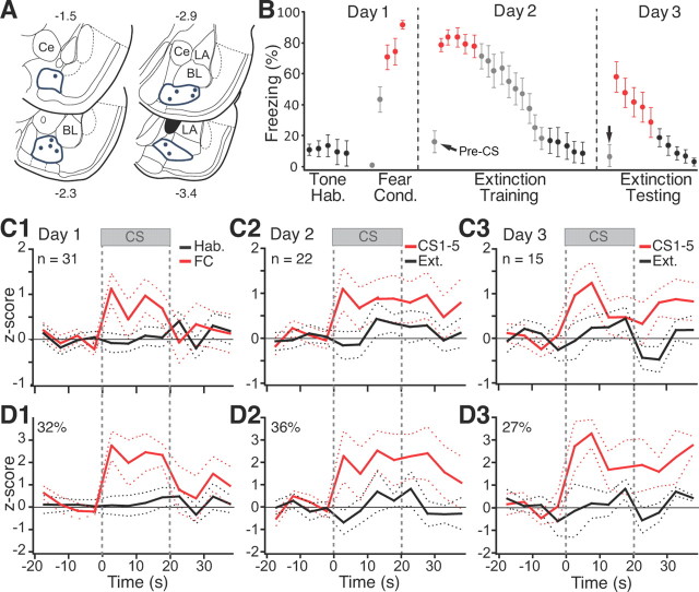
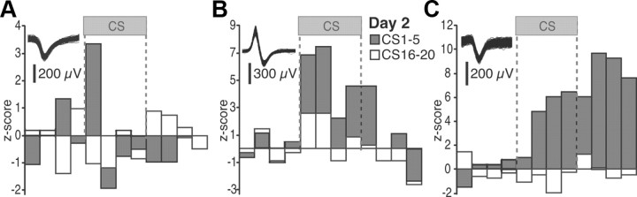
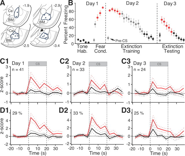
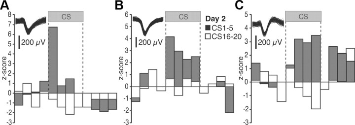
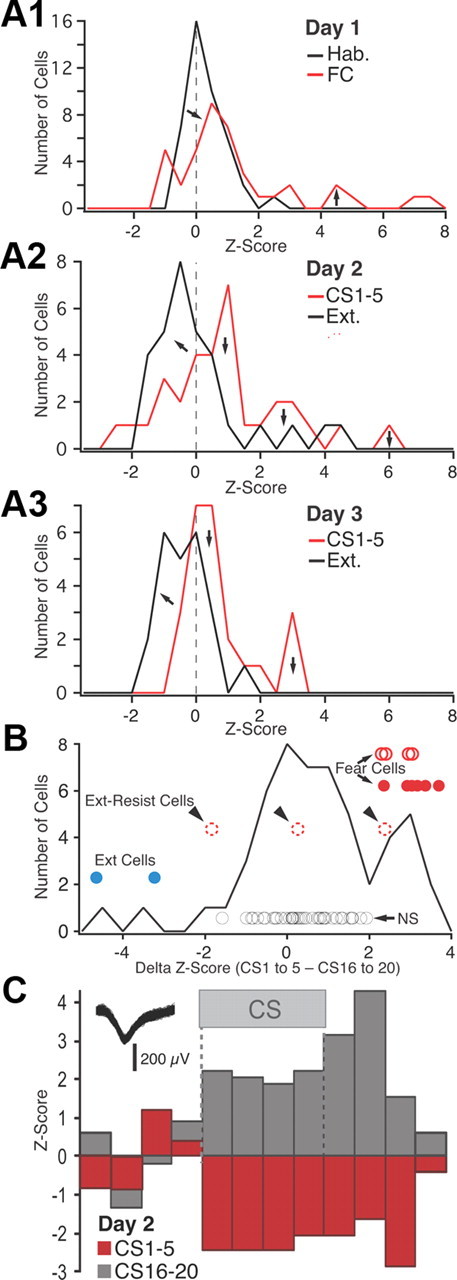
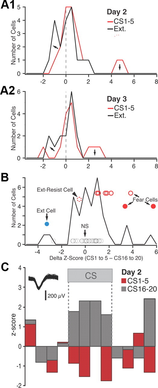
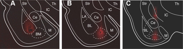
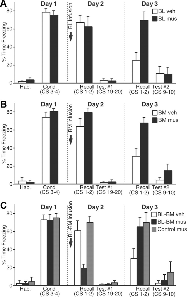
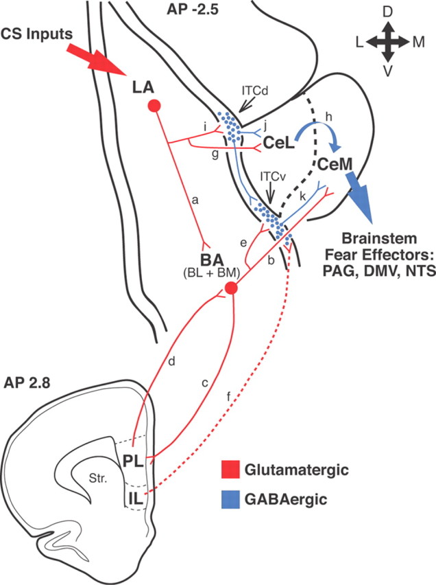
The lateral nucleus (LA) is the input station of the amygdala for information about conditioned stimuli (CSs), whereas the medial sector of the central nucleus (CeM) is the output region that contributes most amygdala projections to brainstem fear effectors. However, there are no direct links between LA and CeM. As the main target of LA and with its strong projection to CeM, the basomedial amygdala (BM) constitutes a good candidate to bridge this gap. Consistent with this notion, it was reported that combined posttraining lesions of the basal nuclei [BM plus basolateral nucleus (BL)] abolish conditioned fear responses, whereas selective BL inactivation does not. Thus, we examined the relative contribution of BM and BL to conditioned fear using unit recordings and inactivation with muscimol microinfusions in rats. Approximately 30% of BM and BL neurons acquired robust responses to auditory CSs predicting footshocks. While most BL cells stopped firing at CS offset, BM responses typically outlasted the CS by ≥ 40 s, paralleling the persistence of conditioned fear after the CS. This observation suggests that BM neurons are not passive relays of rapidly adapting LA inputs about the CS. Surprisingly, independent inactivation of either BM or BL with muscimol did not cause a reduction of conditioned freezing even though an extinction recall deficit was seen the next day. In contrast, combined BL-BM inactivation did. Overall, there results support the notion that the basal nuclei are involved in conditioned fear expression and extinction but that there is functional redundancy between them.


Figures

Figure 1.
Figure 1.
CS responsiveness of BM neurons. A, Locations of microwire bundles. B, Behavior of rats used for BM recordings. Hab., Habituation; Cond., conditioning. Error bars indicate SEM. C1–C3, Average CS responses of all cells recorded on days 1–3, respectively. D1–D3, Average restricted to cells with significant increases in firing rate during ≥1 bin of the CS (threshold, ±2.5z).
Figure 2.
Figure 2.
Representative examples of individual BM units. A–C illustrate three different cells. The insets show action potential waveforms for each cell. Firing rates are z scored to the baseline (pre-CS) period (5 s bins). The gray and white bars represent the average of the first and last five CSs of the recall test, respectively.
Figure 3.
Figure 3.
CS responsiveness of BL neurons. A, Locations of microwire bundles. B, Behavior of rats used for BL recordings. Error bars indicate SEM. C1–C3, Average CS responses of all cells recorded on days 1–3, respectively. D1–D3, Average restricted to cells with significant increases in firing rate during ≥1 bin of the CS (threshold, ±2.5z).
Figure 4.
Figure 4.
Representative examples of individual BL units. A–C illustrate three different cells. The insets show action potential waveforms for each cell. Firing rates are z scored to the baseline (pre-CS) period (5 s bins). The gray and white bars represent the average of the first and last five CSs of the recall test, respectively.
Figure 5.
Figure 5.
Frequency distribution of z-scored CS responsiveness in BL neurons. A, Frequency distributions of z-scored CS responses (based on average of the four 5 s CS bins) on day 1 (A1), day 2 (A2), and day 3 (A3). See legend at top right corner of each panel for explanation of color coding. FC, Fear conditioning; Ext., extinction. B, Frequency distribution of the difference in z scores between the first five and last five CSs of extinction training (data obtained on days 2 and 3 are combined). The circles represent individual cells (see text). Their position with respect to the y-axis has no meaning; different cell subtypes are offset to various degrees for clarity. Blue, Extinction cells with significant negative z scores in response to CS 1–5 and positive z scores in response to the last five CSs. Red circles, Fear cells (filled red circles, fear cells that were inhibited by the end of extinction training; empty red circles with solid line, fear cells that became unresponsive by the end of extinction training; empty red circles with dashed lines, fear cells that maintained a significant positive response to the last 5 CSs). Black circles, Cells with nonsignificant CS responses at all time points. C, Example of an extinction cell.
Figure 6.
Figure 6.
Frequency distribution of z-scored CS responsiveness in BM neurons. A, Frequency distributions of z-scored CS responses (based on average of the four 5 s CS bins) on day 2 (A1) and day 3 (A2). See legend at top right corner of each panel for explanation of color coding. B, Frequency distribution of the difference in z scores between the first five and last five CSs of extinction training (data obtained on days 2 and 3 are combined). The circles represent individual cells that reached significance (see text). Their position with respect to the y-axis has no meaning; different cell subtypes are offset to various degrees for clarity. Blue, Extinction cell with significant negative z scores in response to CS 1–5 and positive z scores in response to CS 16–20. Red circles, Fear cells (filled red circles, fear cells that were inhibited by the end of extinction training; empty red circles with solid line, fear cells that became unresponsive by the end of extinction training; empty red circles with dashed lines, fear cells that maintained a significant positive response to CS 16–20). Black circles, Cells with nonsignificant CS responses at all time points. C, Example of extinction cell.
Figure 7.
Figure 7.
Extent of muscimol diffusion from the infusion site. At each infusion site, the rats received 0.3 μl of 0.5 mm fluorescent muscimol dissolved in aCSF, as in the behavioral experiments. A–C, Muscimol diffusion 10 min after infusion in BL (A), BM (B), or in BM plus dorsal to Ce (C). Ce, Central nucleus of the amygdala; IC, internal capsule; M, medial nucleus of the amygdala; Str, striatum; Th, thalamus.
Figure 8.
Figure 8.
Impact of muscimol infusions in the basal nuclei on fear expression. A–C, CS-elicited freezing (y-axis; average ± SEM) during the behavioral protocol (x-axis). Ten minutes before testing recall, we performed bilateral infusions of vehicle (empty bars) or muscimol (filled bars) in BL (A), BM (B), or both (C). C, Gray bars, Rats that received muscimol in BM plus a site dorsal to Ce (Fig. 7C).
Figure 9.
Figure 9.
Network interactions supporting the acquisition and extinction of conditioned fear responses. Thalamic and cortical afferents convey CS information to LA (large red arrow on top left). CeM constitutes the main source of amygdala projections to brainstem fear effectors (large blue arrow on right). However, LA has no direct connections to CeM neurons. LA can influence CeM indirectly via the basal nuclei (BA) (a, b) or central lateral (CeL) nucleus (g, h). Our results support the notion that in an intact brain, LA can excite CeM neurons via the basal nuclei. However, if the BA nuclei are lesioned before training, conditioning occurs normally, suggesting that CeL might also participate. Disinhibition of CeM from inputs arising in CeL might be involved (Ciocchi et al., 2010; Duvarci et al., 2011). Interactions between LA, BA, and Ce are regulated by intercalated (ITC) cells located at the BLA–Ce border (blue circles) as well as the infralimbic (IL) and prelimbic (PL) regions of the medial prefrontal cortex (bottom scheme). In rats, the medial ITC cells occur in multiple main clusters: one located lateral to CeL (ITCd) and others located more ventrally (ITCv). ITCd receives glutamatergic inputs from LA (i) and project to CeL (j). ITCv receives glutamatergic inputs from BA (e) and project to CeM (k). In addition, ITCd projects to ITCv. After fear conditioning, via LA neurons, the CS excites ITCd neurons more strongly, likely resulting in the inhibition of CeL cells and disinhibition of CeM neurons. The increased recruitment of ITCd cells would also cause the inhibition of ITCv neurons, resulting in further CeM disinhibition. These two disinhibitory mechanisms would reinforce the glutamatergic excitation of CeM by BA inputs. It is likely that the persistence of BM responses after CS offset depends on interactions with the prelimbic cortex (c, d). In contrast, IL would regulate extinction by enabling a potentiation of BA inputs to ITCv neurons (f), resulting in increased feedforward inhibition of CeM cells by ITCv cells (e, k). PAG, Periaqueductal gray; DMV, dorsal motor nucleus of the vagus; NTS, nucleus of the solitary tract.

References

    1. Amano T, Unal CT, Paré D. Synaptic correlates of fear extinction in the amygdala. Nat Neurosci. 2010;13:489–494. - PMC - PubMed
    1. Amorapanth P, LeDoux JE, Nader K. Different lateral amygdala outputs mediate reactions and actions elicited by a fear-arousing stimulus. Nat Neurosci. 2000;3:74–79. - PubMed
    1. Anglada-Figueroa D, Quirk GJ. Lesions of the basal amygdala block expression of conditioned fear but not extinction. J Neurosci. 2005;25:9680–9685. - PMC - PubMed
    1. Burgos-Robles A, Vidal-Gonzalez I, Quirk GJ. Sustained conditioned responses in prelimbic prefrontal neurons are correlated with fear expression and extinction failure. J Neurosci. 2009;29:8474–8482. - PMC - PubMed
    1. Cassell MD, Gray TS, Kiss JZ. Neuronal architecture in the rat central nucleus of the amygdala: a cytological, hodological, and immunocytochemical study. J Comp Neurol. 1986;246:478–499. - PubMed

Publication types

MeSH terms