Case Reports
. 2011 Oct 28:6:104.
doi: 10.1186/1746-1596-6-104.

Peritoneal keratin granuloma associated with endometrioid adenocarcinoma of the uterine corpus

Affiliations
Case Reports

Peritoneal keratin granuloma associated with endometrioid adenocarcinoma of the uterine corpus

Keiichiro Uehara et al. Diagn Pathol. .

Abstract

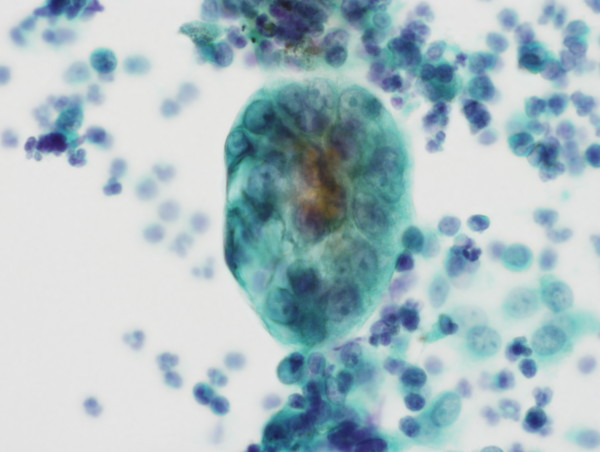
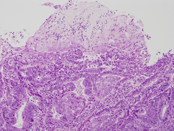
We present a 69-year-old woman with a chief complaint of postmenopausal bleeding. She was diagnosed as having an endometrioid adenocarcinoma by biopsy, and underwent a total abdominal hysterectomy. At the time of surgery, granulation tissue-like nodules were found on the peritoneal serosa of the uterus. In the intraoperative cytology of peritoneal washing, atypical cells were noted. The intraoperative frozen section of the peritoneal nodule revealed granulation tissue with proliferating mesothelial cells. Microscopic examination of the permanent section showed keratin granulomas without viable adenocarcinoma cells on the serosal surface of the ovaries, fallopian tubes and broad ligaments. Postoperative chemotherapy was administered. She has been alive with no evidence of recurrence for 6 months postoperatively. It should be noted that the prognosis of cases in peritoneal keratin granuloma without viable cancer cells is favorable, and that the histological examination is essential for its diagnosis.


Figures

Figure 1
Figure 1
Intraoperative peritoneal washing cytology shows atypical cells with high N/C ratio and conspicuous nucleoli. It is difficult to distinguish between reactive mesothelial cells and adenocarcinomatous cells.
Figure 2
Figure 2
Well differentiated endometrioid adenocarcinoma showing squamous metaplasia. Virtual Slide: http://diagnosticpathology.slidepath.com/dih/webViewer.php?snapshotId=1313178756
Figure 3
Figure 3
Keratin granuloma is composed of eosinophilic laminated keratin surrounded by multinucleated giant cells and chronic inflammatory cells.
Figure 4
Figure 4
Reactive mesothelial cells close to keratin granulomas reveal papillary and gland-like structures.
Figure 5
Figure 5
Immunohistochemically, ghost squamous cells and reactive mesothelial cells are positive for AE1/AE3 in keratin granuloma.
Figure 6
Figure 6
Immunohistochemically, only reactive mesothelial cells near keratin granuloma for calretinin.

References

    1. Chen KT, Kostich ND, Rosai J. Peritoneal foreign body granulomas to keratin in uterine adenoacanthoma. Arch Pathol Lab Med. 1978;102:174–7. - PubMed
    1. Kim KR, Scully RE. Peritoneal keratin granulomas with carcinomas of endometrium and ovary and atypical polypoid adenomyoma of endometrium. A clinicopathological analysis of 22 cases. Am J Surg Pathol. 1990;14:925–32. doi: 10.1097/00000478-199010000-00005. - DOI - PubMed
    1. Montes M, Beautyman W, Haidak GL. Cholesteatomatous endometriosis. Am J Obst & Gynec. 1961;82:119–23. - PubMed
    1. van der Horst C, Evans AJ. Peritoneal keratin granulomas complicating endometrial carcinoma: a report of two cases and review of the literature. Int J Gynecol Cancer. 2008;18:549–53. doi: 10.1111/j.1525-1438.2007.01033.x. - DOI - PubMed
    1. Williams WD, Amazon K, Rywlin AM. Peritoneal keratin globules in uterine adenosquamous carcinoma. South Med J. 1984;77:1316–8. doi: 10.1097/00007611-198410000-00028. - DOI - PubMed

Publication types

MeSH terms