. 2011 Oct 28;30(1):103.
doi: 10.1186/1756-9966-30-103.

Compound Kushen Injection suppresses human breast cancer stem-like cells by down-regulating the canonical Wnt/β-catenin pathway

Affiliations

Compound Kushen Injection suppresses human breast cancer stem-like cells by down-regulating the canonical Wnt/β-catenin pathway

Weiru Xu et al. J Exp Clin Cancer Res. .

Abstract

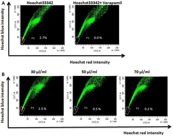
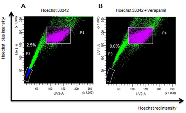
Background: Cancer stem cells (CSCs) play an important role in cancer initiation, relapse and metastasis. To date, no specific medicine has been found to target CSCs as they are resistant to most conventional therapies and proliferate indefinitely. Compound Kushen Injection (CKI) has been widely used for cancer patients with remarkable therapeutic effects in Chinese clinical settings for many years. This study focused on whether CKI could inhibit MCF-7 SP cells in vitro and in vivo.

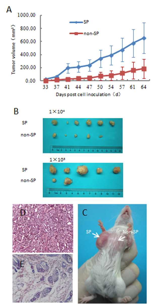
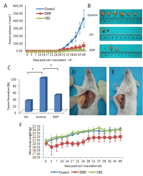
Methods: The analysis of CKI on SP population and the main genes of Wnt signaling pathway were studied first. Then we studied the tumorigenicity of SP cells and the effects of CKI on SP cells in vivo. The mice inoculated with 10,000 SP cells were randomly divided into three groups (6 in each group) and treated with CKI, cisplatin and saline (as a control) respectively for 7 weeks. The tumor formation rates of each group were compared. The main genes and proteins of the Wnt signaling pathway were analyzed by RT-PCR and western blot.

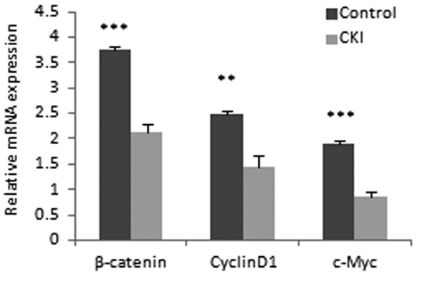
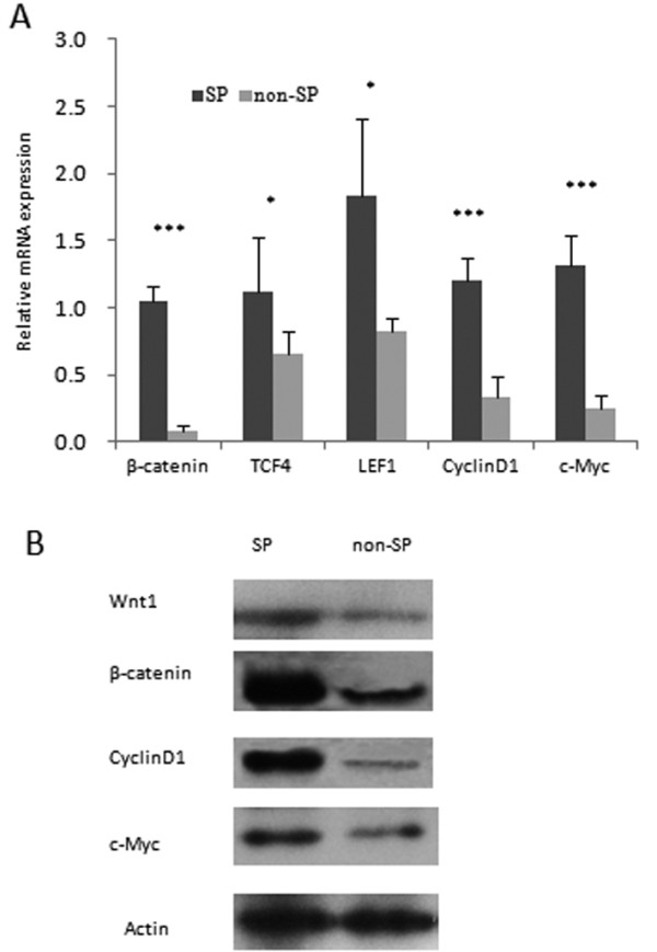
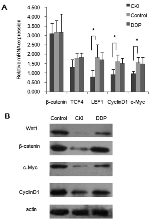
Results: CKI suppressed the size of SP population (approximately 90%), and down-regulated the main genes of Wnt signaling pathway. We also determined that MCF-7 SP cells were more tumorigenic than non-SP and unsorted cells. The Wnt signaling pathway was up-regulated in tumors derived from SP cells compared with that in tumors from non-SP cells. The tumor formation rate of the CKI Group was 33% (2/6, P < 0.05), and that of Cisplatin Group was 50%(3/6, P < 0.05), whereas that of the Control Group was 100% (6/6).The RT-PCR and western blot results indicated that CKI suppressed tumor growth by down-regulating the Wnt/β-catenin pathway, while cisplatin activated the Wnt/β-catenin pathway and might spare SP cells.

Conclusions: It suggested that CKI may serve as a novel drug targeting cancer stem-like cells, though further studies are recommended.


Figures

Figure 1
Figure 1
Analysis of SP cells by CKI treatment. (A) MCF-7 cells were labeled with Hoechst 33342 and analyzed by flow cytometry or with the addition of Verapamil. The percentage of SP cells appeared as the Hoechst low fraction in the P3 is about 2.7%. (B) MCF-7 cells were treated with CKI (30 μl/ml, 50 μl/ml, 70 μl/ml) for 48 h, and SP cells were analyzed by flow cytometry. P3 gate is the percentage of SP cells. Data from a representative experiment (from a total of three) are shown.
Figure 2
Figure 2
The main genes of Wnt/β-catenin pathway was down-regulated in the CKI group in vitro. Quantitative RT-PCR analysis revealed that the expression of β-catenin, CyclinD1 and c-Myc (mean ± SD) were lower in CKI group than those in the control group. Most of the differences were statistically significant (** P < 0.01,*** P < 0.001).
Figure 3
Figure 3
Cell sorting results. MCF-7 cells were labeled with Hoechst 33342 and analyzed by flow cytometry (A) or with the addition of Verapamil (B) SP cells appeared as the Hoechst low fraction in the P3 gate about 2.5%, while non-SP cells retained high levels of Hoechst staining in the P4 gate. Both SP and non-SP cells were sorted, respectively.
Figure 4
Figure 4
SP cells were more tumorigenic. (A) Tumor volumes (mean ± SEM) were plotted for 1 × 10³ cells of each population (SP, non-SP) injected (n = 6 per group). Tumors derived from SP were larger than those from non-SP. (B) Representative tumors due to injection of SP cells (1 × 10⁴ cells, 1 × 10³ cells) compared with non-SP injection (1 × 10⁴ cells, 1 × 10³ cells). (C) A representative tumor in a mouse specimen at the SP injection (1 × 10³ cells) site, but not at the non-SP injection (1 × 10³ cells) site. Histology from the SP injection site ((D), Original magnification, ×200) contained malignant cells, whereas the non-SP injection site ((E), Original magnification, ×200) revealed only normal mammary tissue.
Figure 5
Figure 5
Wnt/β-catenin was up-regulated in tumors derived from SP cells.(A) Quantitative RT-PCR analysis revealed that the expression of β-catenin, TCF4, LEF1, CyclinD1 and c-Myc (mean ± SD) were higher in tumors derived from SP than those in tumors from non-SP. These differences were all statistically significant (* P < 0.05, ***P < 0.001). (B) Western blotting analysis showed that Wnt1, β-catenin, CyclinD1 and c-Myc in tumors derived from SP expressed higher than those in tumors from non-SP cells. The experiment was run in triplicate.
Figure 6
Figure 6
In vivo efficacy of CKI in the MCF-7 SP xenograft model. (A) Tumor volumes (Mean ± SEM) were plotted for each group (n = 6 per group). Both CKI and DDP suppressed tumor growth. (B) A representative comparison image of the incised tumors from CKI, DDP, and the control group. (C) The tumor formation rate of the control group was 100% (6/6), while that of CKI group was 33.33% (2/6) and that of the DDP group was 50% (3/6) (* P < 0.05). (D) A representative mouse specimen without a tumor from the CKI group. (E) A representative specimen with a tumor from the control group. (F) Schematic outline of mice body weight (mean ± SD). No body weight loss was observed in the CKI group, but a slight body weight loss was observed in the DDP group compared to the control group.
Figure 7
Figure 7
The Wnt/β-catenin pathway was down-regulated in the CKI group and up-regulated in the DDP group. a Quantitative RT-PCR analysis revealed that the expression of β-catenin, TCF4, LEF1, CyclinD1 and c-Myc (mean ± SD) were lower in CKI group than those in the control group. Most of the differences were statistically significant (* P < 0.05). The expression of β-catenin, TCF4, LEF1, CyclinD1 and c-Myc (mean ± SD) in DDP group were comparable to those in the control group. b Western blot analysis showed that Wnt1, β-catenin, CyclinD1 and c-Myc in the CKI group were significantly lower than those observed in the control group. The protein level of Wnt1, β-catenin, CyclinD1, and c-Myc in DDP group were comparable to those in the control group. The experiment was run in triplicate.

References

    1. Reya T, Morrison SJ, Clarke MF, Weissman IL. Stem cells, cancer, and cancer stem cells. Nature. 2001;414:105–111. doi: 10.1038/35102167. - DOI - PubMed
    1. Gottesman MM. Mechanisms of cancer drug resistance. Annu Rev Med. 2002;53:615–627. doi: 10.1146/annurev.med.53.082901.103929. - DOI - PubMed
    1. Zhou S, Schuetz JD, Bunting KD, Colapietro AM, Sampath J, Morris JJ, Lagutina I, Grosveld GC, Osawa M, Nakauchi H, Sorrentino BP. The ABC transporter Bcrp1/ABCG2 is expressed in a wide variety of stem cells and is a molecular determinant of the side-population phenotype. Nat Med. 2001;7:1028–1034. doi: 10.1038/nm0901-1028. - DOI - PubMed
    1. Bao S, Wu Q, Mclendon RE, Hao Y, Shi Q, Hjelmeland AB, Dewhirst MW, Bigner DD, Rich JN. Glioma stem cells promote radioresistance by preferential activation of the DNA damage response. Nature. 2006;444:756–760. doi: 10.1038/nature05236. - DOI - PubMed
    1. Graham SM, Jorgensen HG, Allan E, Pearson C, Alcorn MJ, Richmond L, Holyoake TL. Primitive, quiescent, Philadelphia-positive stem cells from patients with chronic myeloid leukemia are insensitive to STI571 in vitro. Blood. 2002;99:319–325. doi: 10.1182/blood.V99.1.319. - DOI - PubMed

Publication types

MeSH terms

LinkOut - more resources