Review
. 2011 Nov;29(4):801-23.
doi: 10.1016/j.ncl.2011.07.014.

Epilepsy and the consciousness system: transient vegetative state?

Affiliations
Review

Epilepsy and the consciousness system: transient vegetative state?

Hal Blumenfeld. Neurol Clin. 2011 Nov.

Abstract

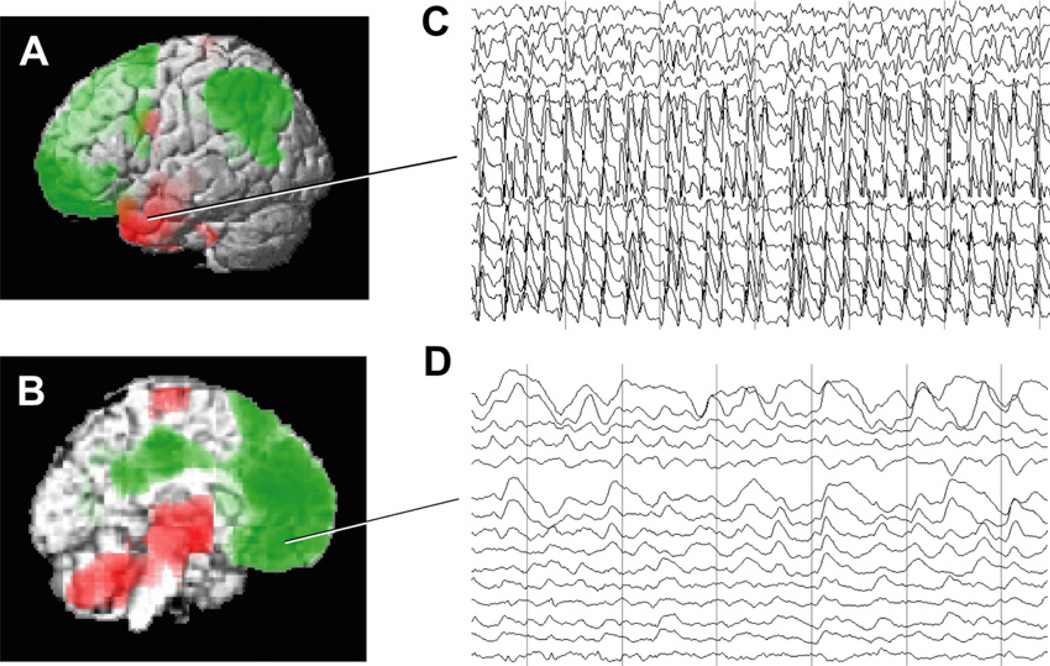
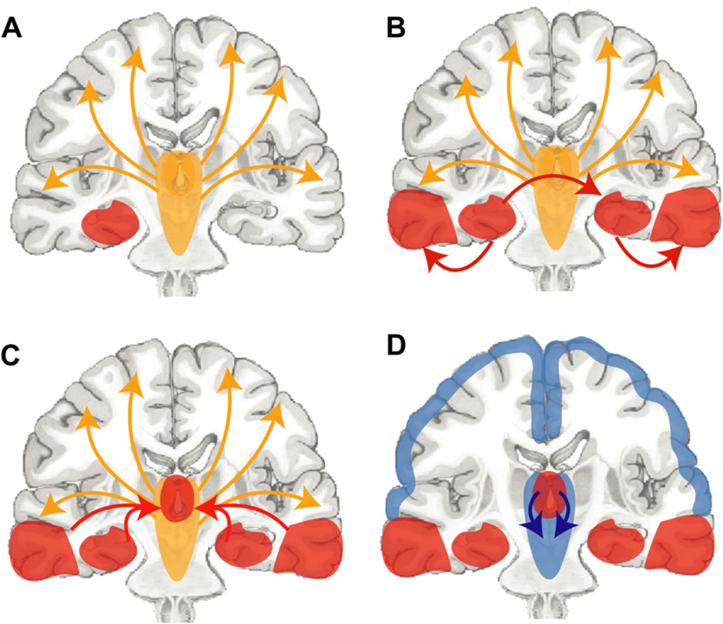
Recent advances have shown much in common between epilepsy and other disorders of consciousness. Behavior in epileptic seizures often resembles a transient vegetative or minimally conscious state. These disorders all converge on the "consciousness system" -the bilateral medial and lateral fronto-parietal association cortex and subcortical arousal systems. Epileptic unconsciousness has enormous clinical significance leading to accidental injuries, decreased work and school productivity, and social stigmatization. Ongoing research to better understand the mechanisms of impaired consciousness in epilepsy, including neuroimaging studies and fundamental animal models, will hopefully soon enable treatment trails to reduce epileptic unconsciousness and improve patient quality of life.


Figures

Fig. 1
Fig. 1
The consciousness system. Anatomic structures involved in regulating the level of alertness, attention, and awareness. (A) Medial view showing cortical (blue) and subcortical (red) components of the consciousness system. (B) Lateral cortical components of the consciousness system. Note that other circuits not pictured here, such as the basal ganglia and cerebellum, may also play a role in attention and other aspects of consciousness. (Reproduced from Blumenfeld H. Neuroanatomy through clinical cases. 2nd edition. Sunderland (MA): Sinauer Associates; 2010; with permission.)
Fig. 2
Fig. 2
Electroencephalography (EEG) and behavior during typical absence seizures. (A) EEG showing typical 3- to 4-Hz spike-wave discharge during an absence seizure. Amplitude is maximal in frontal electrodes (FP1, FP2, F7, F3, FZ, F4, F8) and lower in more posterior occipital regions (O1, O2). A series of letters were presented to the patient (Stimuli) in a continuous performance task. Prior to the seizure the patient pushed the button (Response, voltage deflections) correctly to each target letter (X). However, when the target letter X occurred during the seizure, the patient was unable to respond. Linked-ears referential EEG recording. Functional magnetic resonance imaging (fMRI) changes for this seizure are shown in Fig. 3. (B) Average behavioral impairment during absence seizures. Percent correct responses are shown over time (2-second time bins) before, during, and after seizures (shaded region, normalized to mean seizure duration of ~6 seconds). Results are shown for 2 different tasks: in the continuous performance task (CPT) random letters appeared once per second and patients were instructed to push a button each time the target letter X appeared (see also A); in the repetitive tapping task (RTT) patients were instructed to push the button for every letter regardless of its identity. Performance on the more difficult CPT task declined rapidly for letters presented just before seizure onset and recovered quickly after seizure end. Impaired performance on the RTT task was more transient than on CPT, did not begin until after seizure onset, and was less severely impaired during seizures than the CPT task (F = 15.3, P = .017; analysis of variance). Results are based on a total of 53 seizures in 8 patients; 41 seizures in 5 patients during CPT and 12 seizures in 4 patients during RTT. ([A] Reproduced from Berman R, Negishi M, Vestal M, et al. Simultaneous EEG, fMRI, and behavioral testing in typical childhood absence seizures. Epilepsia 2010;51(10):2011–22; with permission; and [B] Bai X, Vestal M, Berman R, et al. Dynamic time course of typical childhood absence seizures: EEG, behavior, and functional magnetic resonance imaging. J Neurosci 2010;30:5884–93; with permission.)
Fig. 3
Fig. 3
fMRI changes during a typical absence seizure involve the consciousness system and primary cortices. Blood oxygen level dependent (BOLD) fMRI changes are shown from a 12-second seizure in a 14-year-old girl with childhood absence epilepsy (EEG for this seizure is shown in Fig. 2A). The consciousness system demonstrates BOLD fMRI increases in the thalamus, decreases in the interhemispheric regions (anterior cingulate, precuneus), decreases in the lateral parietal cortex, and increases in the lateral frontal cortex. In addition, fMRI increases are present in the primary cortices including the primary visual (occipital), primary auditory (superior temporal), and primary sensorimotor (Rolandic) cortex. fMRI decreases are also seen in the pons, basal ganglia, and cerebellum. Results were analyzed in SPM2 (http://www.fil.ion.ucl.ac.uk/SPM) using a t-test to compare seizure versus baseline with uncorrected height threshold (P = .001) and extent threshold (k = 3 voxels). (Unpublished data Courtesy of R. Berman).
Fig. 4
Fig. 4
Generalized tonic-clonic seizure induced by bilateral frontotemporal electroconvulsive therapy. Ictal single-photon emission computed tomography (SPECT) image for a single generalized tonic-clonic seizure compared with interictal baseline (red = cerebral blood flow [CBF] increases; green = CBF decreases). (A) Lateral view. (B) Medial view. Changes in the consciousness system include CBF increases in lateral frontotemporal cortex, lateral parietal and medial parietal cortex. CBF increases were also present in the thalamus (best seen in cross sections, not shown), as well as in the cerebellum. CBF decreases were present in the medial frontal and cingulate cortex, as well as in lateral cortical regions. SPM extent threshold k = 125 voxels; height threshold: P = .01. (Modified from Blumenfeld H, McNally KA, Ostroff RB, et al. Targeted prefrontal cortical activation with bifrontal ECT. Psychiatry Res 2003;123:165–70; with permission.)
Fig. 5
Fig. 5
Frontoparietal CBF decreases and thalamic increases are correlated with increased CBF in the cerebellum during and following generalized tonic-clonic seizures. Network correlations are shown for spontaneous secondarily generalized tonic-clonic seizures imaged with ictal SPECT in epilepsy patients during video/EEG monitoring. Positive (red) and negative (green) correlations are shown between CBF changes in the cerebellum and the rest of the brain across patients (n = 59 seizures in 53 patients). Significant positive correlations were found between the cerebellum and the upper brainstem tegmentum and thalamus. Negative correlations were found with the bilateral frontoparietal association cortex, anterior and posterior cingulate, and precuneus. Images were analyzed with SPM extent threshold k = 125 voxels, and height threshold P = .01. (Reproduced from Blumenfeld H, Varghese G, Purcaro MJ, et al. Cortical and subcortical networks in human secondarily generalized tonic-clonic seizures. Brain 2009;132:999–1012; with permission.)
Fig. 6
Fig. 6
CBF and EEG changes in temporal lobe complex partial seizures. (A, B) Group analysis of SPECT ictal-interictal difference imaging during temporal lobe seizures. CBF increases are present in the temporal lobe (A) and in the medial thalamus (B). Decreases are seen in the lateral frontoparietal association cortex (A) and in the interhemispheric regions (B). (C, D) Intracranial EEG recordings from a patient during a temporal lobe seizure. High-frequency polyspike-and-wave seizure activity is seen in the temporal lobe (C). The orbital and medial frontal cortex (and other regions, EEG not shown) do not show polyspike activity, but instead large-amplitude, irregular slow rhythms resembling coma or sleep (D). Vertical lines in C and D denote 1-second intervals. Note that the EEG and SPECT data were from similar patients, but were not simultaneous, and are shown together here for illustrative purposes only. ([A, B] Modified from Blumenfeld H, McNally KA, Vanderhill SD, et al. Positive and negative network correlations in temporal lobe epilepsy. Cerebral Cortex 2004;14:892–902; with permission; and [C, D] Blumenfeld H, Rivera M, McNally KA, et al. Ictal neocortical slowing in temporal lobe epilepsy. Neurology 2004;63:1015–21; with permission.)
Fig. 7
Fig. 7
Network inhibition hypotheses for impaired consciousness during temporal lobe complex partial seizures. (A) Under normal conditions, the upper brainstem-diencephalic activating systems interact (yellow arrows) with the cerebral cortex to maintain normal consciousness. A focal seizure involving the mesial temporal lobe begins unilaterally (red region). If it remains unilateral then a simple-partial seizure will occur without impairment of consciousness. (B) Propagation (red arrows) of seizure activity from the mesial temporal lobe to the ipsilateral lateral temporal lobe and the contralateral temporal lobe. (C). Spread of seizure activity from bilateral temporal lobes to midline subcortical structures. (D). Inhibition (blue arrows) of the midline subcortical structures, together with the resulting depressed activity in bilateral frontoparietal association cortex in complex-partial seizures, leads to loss of consciousness. (Reproduced from Englot DJ, Yang L, Hamid H, et al. Impaired consciousness in temporal lobe seizures: role of cortical slow activity. Brain 2010;133(Pt 12): 3764–77; with permission.)

References

    1. Plum F, Posner JB. The diagnosis of stupor and coma. 3rd edition. Philadelphia: Davis; 1982.
    1. Blumenfeld H. Neuroanatomy through clinical cases. Sunderland (MA): Sinauer Assoc. Publ., Inc; 2002.
    1. Blumenfeld H. Epilepsy and consciousness. Chapter 2. In: Laureys S, editor. The neurology of consciousness: cognitive neuroscience and neuropathology. New York: G Tononi Academic Press; 2009. pp. 15–30.
    1. Blumenfeld H. Neuroanatomy through clinical cases. 2nd edition. Sunderland (MA): Sinauer Assoc Publ Co; 2010.
    1. Steriade M, Jones EG, McCormick DA, editors. Thalamus. Amsterdam: Elsevier Science; 1997.

Publication types