Review
. 2012 Jan;33(1):17-27.
doi: 10.1016/j.tips.2011.09.003. Epub 2011 Oct 25.

Diversity and modularity of G protein-coupled receptor structures

Affiliations
Review

Diversity and modularity of G protein-coupled receptor structures

Vsevolod Katritch et al. Trends Pharmacol Sci. 2012 Jan.

Abstract

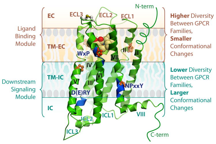
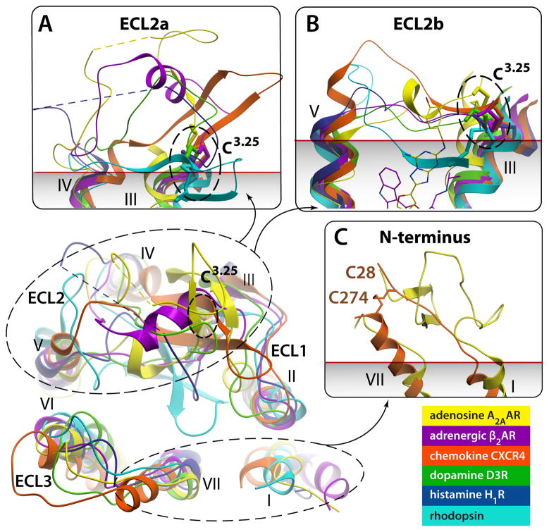
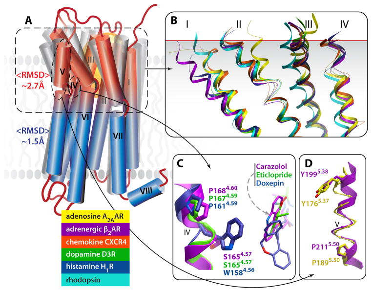
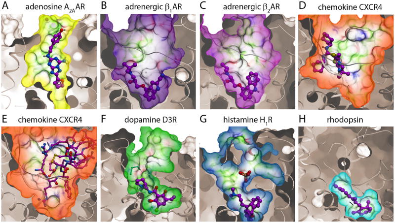
G protein-coupled receptors (GPCRs) comprise the most 'prolific' family of cell membrane proteins, which share a general mechanism of signal transduction, but greatly vary in ligand recognition and function. Crystal structures are now available for rhodopsin, adrenergic, and adenosine receptors in both inactive and activated forms, as well as for chemokine, dopamine, and histamine receptors in inactive conformations. Here we review common structural features, outline the scope of structural diversity of GPCRs at different levels of homology, and briefly discuss the impact of the structures on drug discovery. Given the current set of GPCR crystal structures, a distinct modularity is now being observed between the extracellular (ligand-binding) and intracellular (signaling) regions. The rapidly expanding repertoire of GPCR structures provides a solid framework for experimental and molecular modeling studies, and helps to chart a roadmap for comprehensive structural coverage of the whole superfamily and an understanding of GPCR biological and therapeutic mechanisms.


Figures

Fig. 1
Fig. 1
Current status of GPCR family structural coverage. Published high-resolution structures of GPCRs are shown on the sequence homology tree (modified from [2]). Highlighted areas show close homologs of the crystal structures with better than 35% sequence identity in the TM helices, which are likely to be amenable for accurate comparative modeling.
Fig. 2
Fig. 2
General architecture and modularity of GPCRs. Major regions and structural features of GPCRs are shown on example of the D3R crystal structure (PDB ID 3PBL). The EC region includes three ECLs and the N-terminus, which may be represented by a short unstructured peptide (as in most class A GPCRs), or a longer globular-like domain. The 7TM helical bundle contains a number of proline-dependent kinks (prolines shown in orange), that approximately divide the receptor into two modules. The EC module (EC and TM-EC regions) is responsible for binding diverse ligands and has much higher structural diversity. In contrast, the IC module (IC and IC-TM regions), involved in binding downstream effectors including G proteins and arrestins, is more conserved between GPCRs, but undergoes larger conformational changes upon receptor activation. Blue ribbon patches highlight highly conserved, functionally relevant motifs in the TM helices of Class A GPCRs. The C-terminus in most GPCRs is comprised of helix VIII, and in many receptors also has a palmitoylation site anchoring helix VIII to the membrane (not shown).
Fig. 3
Fig. 3
Remarkable diversity in the EC region. Top view of ECLs from superimposed GPCR structures with close-up side views in panels (A)–(C). Note different secondary structure elements (or lack thereof) in ECL2, as well as large deviations in positions of extracellular tips of the TM helices. N-termini of Rhodopsin and CXCR4 are removed for clarity. (A) Close up of ECL2a, a part of ECL2 from helix IV to the conserved disulfide bond connecting ECL2 to helix III. The ECL2a is highly variable in length and secondary structure elements, showing distinct structure between structures of all GPCRs solved; the only exception being the practically identical ECL2a conformations ((RMSD(Cα)=0.5 A)) between very closely related β2AR (PDB ID 2RH1) and β1AR (PDB ID 2VT4, not shown). Short unresolved portions of ECL2a in A2AAR and H1R are shown by dashed lines. (B) Close up side view of ECL2b – the linker part connecting helices V and III through the disulfide bond to Cys3.25. Note that in some GPCRs the ECL2b element is as short as 4–5 amino acids and fully extended, while longer sequence in others can comprise small secondary structure elements. Relative positions of Carazolol and ZM241385 interacting with ECL2b residues are shown by thin lines. (C) N-terminus is fully resolved in Rhodopsin (PDB ID 1GZM; residues 1:34) and partially in CXCR4 (PDB ID 3ODU; residues 27:34) structures; in other known structures N-termini are likely disordered. Approximate membrane boundary is shown in panels (A)–(C), as predicted by OPM database [86].
Fig. 4
Fig. 4
Structural diversity and conformational plasticity in GPCRs. (A) A cartoon illustrating 7TM helical bundles of known GPCR structures. In general, structural deviations between receptors are larger in TM-EC module (red) than in TM-IC module (blue) (average RMSDs are ~2.7 A vs. 1.5 A). (B) Structural superimposition of TM helical bundle structures of different GPCR shows especially high deviations in the extracellular half of the bundle, with strong deviation observed in each of the helices (only helices I to IV are shown for clarity). (C) An insertion/deletion in structural alignment, shown in an example of a conserved proline kink in helix IV. Note that Trp in position 4.56 of H1R structurally aligns with Ser in position 4.57 of β2AR and D3R, while the Pro in position 4.60 of β2AR structurally aligns with Pro in position 4.59 in D3R and H1R. (D) Observed π-helical structure in the extracellular portion of A2AAR helix V. The π-helix (i+5) has more residues per helical turn than standard α-helical (i+4) structure, resulting in “phase shift” and structural alignment of Tyr in position 5.37 of A2AAR with Tyr in position 5.38 of β2AR.
Fig. 5
Fig. 5
Diversity of the ligand binding pocket shape and properties in GPCR crystal structures. Structural diversity, including large backbone deviations of ECLs and TM helices, results in dramatic variations in size, shape and binding properties of GPCR pockets: (A) In A2AAR (PDB ID 3EML), the pocket forms an accessible channel with the ligand (antagonist ZM241385) positioned vertically towards the EC region. (B) (C) The β1AR (PDB ID 2VT4) and β2AR (PDB ID 2RH1) have almost identical highly accessible pockets that share the same contact residues for antagonists cyanopindolol and carazolol, respectively. (D) (E) The chemokine CXCR4 receptor has a large and open pocket, which can accommodate not only small molecules such as IT1t (D) (PDB ID 3ODU), but also molecules as large as the 16-residue cyclic peptide CVX15 (E) (PDB ID 3OE0). (F) The D3R pocket (PDB ID 3PBL) has distinct EC and “core” sub-pockets, the latter one occupied by the antagonist eticlopride (see also Fig 6B). (G) The H1R pocket (PDB ID 3RZE) reaches deeper into the TM than in other receptors, while the PO43− ion modulates ligand access to the pocket. (H) The retinal pocket in Rhodopsin (PDB ID 1GZM) is small, hydrophobic and completely enclosed. All pockets are shown in the same orientation.
Fig. 6
Fig. 6
Examples of allosteric interaction sites identified in GPCR crystal structures. A) Binding of a phosphate ion in the ECLs of H1R (PDB ID 3RZE) is coordinated by four receptor side chains, and affects the accessibility of the ligand pocket. This site is key for H1 subtype selectivity to second-generation antihistamines [18]. B) The ligand-binding pocket in the D3R (PDB ID 3PBL) consists of a core site (dark green) with bound antagonist eticlopride and a secondary or allosteric site (lime). Docking the D3 subtype-selective ligand R22 into the D3R crystal structure reveals the bitopic nature of this compound occupying the core site and extending into the secondary site. Interactions between R22 and the secondary site define most of the subtype selectivity for this ligand [17]. C) The cholesterol binding site on the lipid-TM interface has been reproduced in different crystal forms of the β2AR, (shown here by cyan sticks for PDB IDs 2RH1 and by green sticks for 3D4S), and is likely conserved in many other GPCRs [23].

References

    1. Nordstrom KJ, et al. Independent HHsearch, Needleman-Wunsch-based and motif analyses reveals the overall hierarchy for most of the G protein-coupled receptor families. Mol Biol Evol. 2011;28(9):2471–80. - PubMed
    1. Fredriksson R, et al. The G-protein-coupled receptors in the human genome form five main families. Phylogenetic analysis, paralogon groups, and fingerprints. Mol Pharmacol. 2003;63(6):1256–72. - PubMed
    1. Fredriksson R, Schioth HB. The repertoire of G-protein-coupled receptors in fully sequenced genomes. Mol Pharmacol. 2005;67(5):1414–25. - PubMed
    1. Strotmann R, et al. Evolution of GPCR: change and continuity. Mol Cell Endocrinol. 2011;331(2):170–8. - PubMed
    1. Lagerstrom MC, Schioth HB. Structural diversity of G protein-coupled receptors and significance for drug discovery. Nat Rev Drug Discov. 2008;7(4):339–57. - PubMed

Publication types