. 2011 Dec 15;10(24):4208-16.
doi: 10.4161/cc.10.24.18487. Epub 2011 Dec 15.

Energy transfer in "parasitic" cancer metabolism: mitochondria are the powerhouse and Achilles' heel of tumor cells

Affiliations

Energy transfer in "parasitic" cancer metabolism: mitochondria are the powerhouse and Achilles' heel of tumor cells

Ubaldo E Martinez-Outschoorn et al. Cell Cycle. .

Abstract

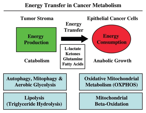
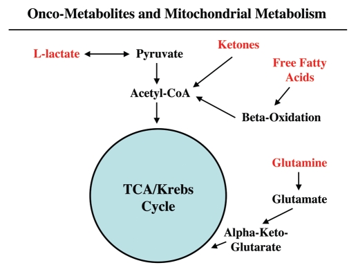
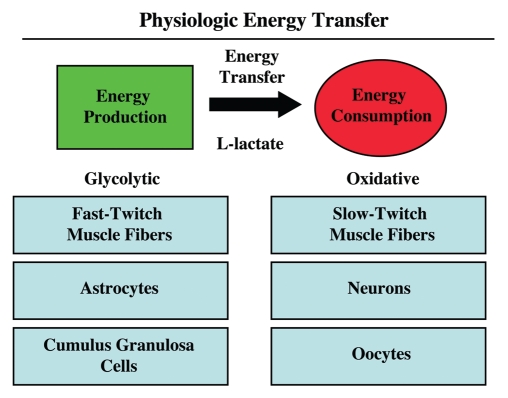
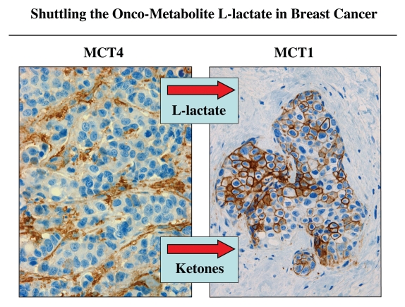
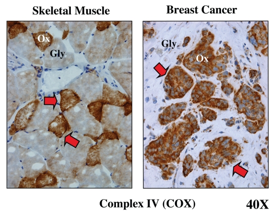
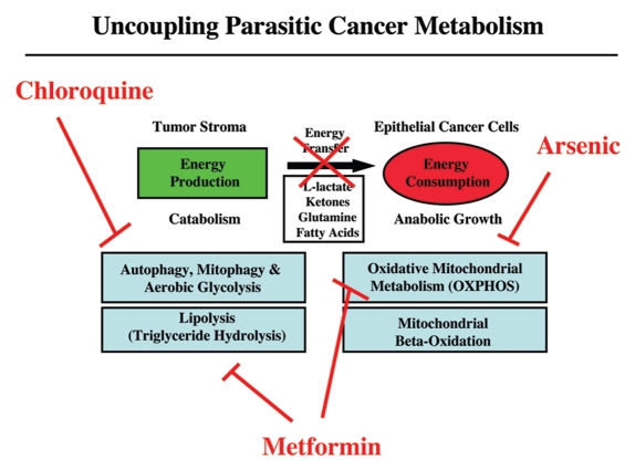
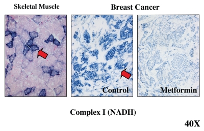
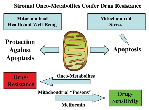
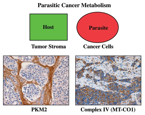
It is now widely recognized that the tumor microenvironment promotes cancer cell growth and metastasis via changes in cytokine secretion and extracellular matrix remodeling. However, the role of tumor stromal cells in providing energy for epithelial cancer cell growth is a newly emerging paradigm. For example, we and others have recently proposed that tumor growth and metastasis is related to an energy imbalance. Host cells produce energy-rich nutrients via catabolism (through autophagy, mitophagy, and aerobic glycolysis), which are then transferred to cancer cells to fuel anabolic tumor growth. Stromal cell-derived L-lactate is taken up by cancer cells and is used for mitochondrial oxidative phosphorylation (OXPHOS) to produce ATP efficiently. However, "parasitic" energy transfer may be a more generalized mechanism in cancer biology than previously appreciated. Two recent papers in Science and Nature Medicine now show that lipolysis in host tissues also fuels tumor growth. These studies demonstrate that free fatty acids produced by host cell lipolysis are re-used via beta-oxidation (beta-OX) in cancer cell mitochondria. Thus, stromal catabolites (such as lactate, ketones, glutamine and free fatty acids) promote tumor growth by acting as high-energy onco-metabolites. As such, host catabolism, via autophagy, mitophagy and lipolysis, may explain the pathogenesis of cancer-associated cachexia and provides exciting new druggable targets for novel therapeutic interventions. Taken together, these findings also suggest that tumor cells promote their own growth and survival by behaving as a "parasitic organism." Hence, we propose the term "Parasitic Cancer Metabolism" to describe this type of metabolic coupling in tumors. Targeting tumor cell mitochondria (OXPHOS and beta-OX) would effectively uncouple tumor cells from their hosts, leading to their acute starvation. In this context, we discuss new evidence that high-energy onco-metabolites (produced by the stroma) can confer drug resistance. Importantly, this metabolic chemo-resistance is reversed by blocking OXPHOS in cancer cell mitochondria with drugs like Metformin, a mitochondrial "poison." In summary, parasitic cancer metabolism is achieved architecturally by dividing tumor tissue into at least two well-defined opposing "metabolic compartments:" catabolic and anabolic.


Figures

Figure 1
Figure 1
Energy transfer in cancer metabolism. The tumor stroma generates catabolites that are transferred to cancer cells for anabolic growth. The stroma has high levels of autophagy, mitophagy, glycolysis and lipolysis, while epithelial cancer cells have high mitochondrial mass and activity (oxidative phosphorylation and β oxidation). Reproduced and modified with permission from reference .
Figure 2
Figure 2
Onco-metabolites derived from the tumor stroma promote anabolic cancer cell growth via the TCA cycle and oxidative mitochondrial metabolism. Note that various stromally derived onco-metabolites (L-lactate, ketones, free fatty acids and glutamine; shown in red) all feed into the TCA/Krebs cycle via either Acetyl-CoA or Alpha-Keto-Glutarate, promoting oxidative mitochondrial metabolism (OXPHOS) in epithelial cancer cells. The end result is highly efficient ATP production in aggressive cancer cells.
Figure 3
Figure 3
Physiologic energy transfer. Metabolic-coupling is a normal and widespread physiological phenomenon that is required to maintain homeostasis or energy balance. Metabolic-coupling occurs in organ systems throughout the body, including skeletal muscle, the brain and the ovary. In all three tissues, a “lactateshuttle” exists. In this context, glycolytic cells (fast-twitch muscle fibers, astrocytes and cumulus/granulosa cells) are metabolically coupled to oxidative cells (slow-twitch muscle fibers, neurons and oocytes). L-lactate is generated in glycolytic cells from glucose and is transferred to oxidative cells, which is efficiently used to make large amounts of ATP, via oxidative mitchondrial metabolism. Monocarboxylate transporters (MCTs) shuttle lactate from one cell type to another.
Figure 4
Figure 4
Shuttling the onco-metabolite L-lactate from the tumor stroma to epithelial cancer cells. Previously, we proposed that a “lactate shuttle” also exists in human breast cancers. More specifically, the distribution of lactate transporters is highly compartmentalized in human breast cancers. Note that MCT4 (a marker of aerobic glycolysis and L-lactate secretion) is largely confined to cancer-associated fibroblasts in the tumor stroma. Conversely, MCT1 (a marker of L-lactate uptake) is localized to epithelial cancer cells. Because of their broad specificity, the same MCT transporters can also function in the shuttling of ketone bodies from the tumor stroma to epithelial cancer cells. Arrows indicate the direction of energy transfer. Reproduced and modified with permission from reference .
Figure 5
Figure 5
Metabolic compartmentalization of mitochondrial activity in skeletal muscle and human breast cancer. Frozen sections from skeletal muscle tissue or human breast cancers were subjected to a routine histochemical stain that detects the functional activity of mitochondrial complex IV [cytochrome C oxidase (COX)]. This allows the visualization of oxidative mitochondrial metabolism in tissue sections. COX-positive cells are positively stained brown (see red arrows). In skeletal muscle, note that fast-twitch fibers are glycolytic (Gly) and are COX-negative, while slow-twitch fibers are oxidative (Ox) and are COX-positive. In breast cancers, the tumor stroma is glycolytic (Gly) and is COX-negative, while epithelial tumor cell nests are oxidative (Ox) and are COX-positive. These results support the idea that tumors show metabolic compartmentalization and specialization, as occurs in skeletal muscle tissue. Reproduced and modified with permission from reference .
Figure 6
Figure 6
Uncoupling parasitic cancer metabolism. Drugs such as chloroquine (which inhibits autophagy) and metformin (which inhibits lipolysis), will prevent energy transfer to cancer cells and tumor growth. In this scenario, mitochondrial poisons (such as metformin, arsenic and others) could also be used to uncouple tumor cells from the energy-producing host stroma.
Figure 7
Figure 7
Visualizing the anti-mitochondrial effects of Metformin in human breast cancer tumor tissue. Frozen sections from human breast cancers were subjected to mitochondrial Complex I (NADH) activity staining. NADH-positive cells are positively stained blue (see red arrows). In breast cancers, note that the tumor stroma is glycolytic and is NADH-negative, while epithelial tumor cell nests are oxidative and are NADH-positive. Note that treatment with Metformin, a known complex I inhibitor, prevented the NADH-staining of epithelial cancer cell nests. Positive controls with skeletal muscle were performed in parallel to ensure the specificity of the staining procedure and allowed us to detect complex I-positive muscle fibers (blue), which represent oxidative slow-twitch fibers. Reproduced and modified with permission from reference .
Figure 8
Figure 8
Stromal onco-metabolites confer drug resistance by promoting mitochondrial health or well-being, providing an escape from stress-induced apoptosis. Simple stromally derived metabolites (such as L-lactate, ketones and glutamine) promote mitochondrial “health” in cancer cells, effectively shutting off their apoptotic machinery, resulting in protection against cell death, even when challenged with anticancer drugs. In contrast, we can overcome metabolite-induced drug resistance in cancer cells by using mitochondrial poisons (such as Metformin).
Figure 9
Figure 9
Metabolic compartments in parasitic cancer metabolism. In summary, we believe that cancer cells act as metabolic parasites and extract nutrients from host cells by inducing catabolic processes (autophagy, mitophagy, aerobic glycolysis and lipolysis). As a consequence, the tumor stroma shows a shift toward aerobic glycolysis, and epithelial cancer cells show functional hyper-activation of oxidative mitochondrial metabolism (OXPHOS). In support of this model, cancer-associated fibroblasts and the tumor stroma overexpress PKM2 (a rate-limiting glycolytic enzyme, left panel). Conversely, breast cancer epithelial cells upregulate MT-CO1 (a key component of mitochondrial complex IV, right panel). The metabolic compartmentalization of PKM2 and MT-CO1 were visualized by immunostaining with specific antibody probes (brown reaction product). Reproduced and modified with permission from references and .

References

    1. Rønnov-Jessen L, Bissell MJ. Breast cancer by proxy: can the microenvironment be both the cause and consequence? Trends Mol Med. 2009;15:5–13. doi: 10.1016/j.molmed.2008.11.001. - DOI - PMC - PubMed
    1. Kenny PA, Lee GY, Bissell MJ. Targeting the tumor microenvironment. Front Biosci. 2007;12:3468–3474. doi: 10.2741/2327. - DOI - PMC - PubMed
    1. Paget S. The distribution of secondary growths in cancer of the breast. Lancet. 1889;133:571–573. doi: 10.1016/S0140-6736(00)49915-0. - DOI - PubMed
    1. Hart IR. ‘Seed and soil’ revisited: mechanisms of site-specific metastasis. Cancer Metastasis Rev. 1982;1:5–16. doi: 10.1007/BF00049477. - DOI - PubMed
    1. Hart IR, Fidler IJ. Role of organ selectivity in the determination of metastatic patterns of B16 melanoma. Cancer Res. 1980;40:2281–2287. - PubMed

Publication types