

Postmortem studies in Parkinson's disease
- PMID: 22033507
- PMCID: PMC3181805
- DOI: 10.31887/DCNS.2004.6.3/ahartmann
Postmortem studies in Parkinson's disease
Abstract
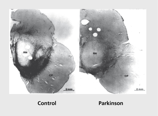
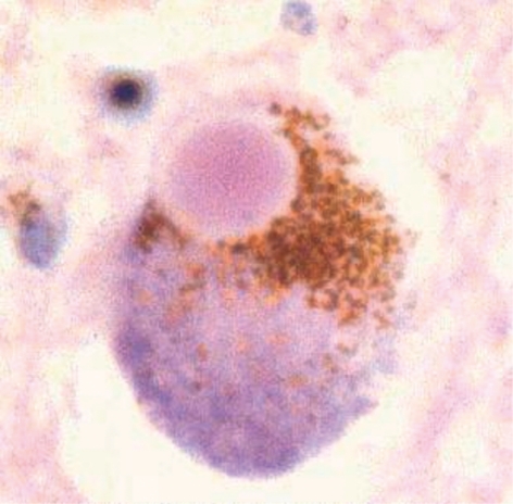
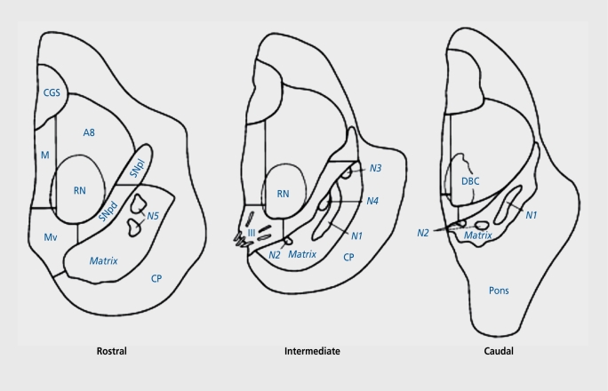
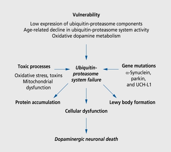
No animal model to date perfectly replicates Parkinson's disease (PD) etiopathogenesis, and the anatomical organization of the nigrostriatal system differs considerably between species. Human postmortem material therefore remains the gold standard for both formulating hypotheses for subsequent testing in in vitro and in vivo PD models and verifying hypotheses derived from experimental PD models with regard to their validity in the human disease. This article focuses on recent and relevant fields in which human postmortem work has generated significant impact in our understanding of PD. These fields include Lewy body formation, regional vulnerability of dopaminergic neurons, oxidative/nitrative cellular stress, inflammation, apoptosis, infectious and environmental agents, and nondopaminergic lesions.
A la fecha, ningún modelo animal ha podido reproducir perfectamente la etiopatogenia de la enfermedad de Parkinson (EP). Además, la organización anatómica del sistema nigroestriatal difiere considerablemente entre las especies. Es por esto que los estudios postmortem en humanos continúan siendo el gold standard tanto para la formulación de hipótesis que serán probadas posteriormente en modelos de EP in vivo e in vitro, como también para la verificación de hipótesis que deriven de modelos experimentales de EP respecto a su validez para la enfermedad en el hombre. Este artículo se centra en campos recientes y relevantes en los que el trabajo postmortem en humanos ha generado un impacto significativo para nuestra comprensión de la EP. Estos campos incluyen la formación de cuerpos de Lewy, la vulnerabilidad regional de las neuronas dopaminérgicas, el estrés celular de la oxidación y de la nitratación, la inflamación, la apoptosis, los agentes infecciosos y ambientales, y las lesiones no dopaminérgicas.
Aucun modèle animal à ce jour ne reproduit fidèlement l'étiopathogenèse de la maladie de Parkinson (MP). Aussi, l'organisation anatomique du système nigrostrié varie considérablement d'une espèce à l'autre. C'est pourquoi les études post mortem chez l'homme restent la meilleure approche soit pour formuler des hypothèses qui seront ensuite testées dans des modèles in vitro et in vivo de la MP, soit pour vérifier des hypothèses dérivées de modèles expérimentaux de la MP, quant à leur validité en pathologie humaine. Dans cet article, l'accent sera mis sur des domaines de recherche récents et pertinents, tels que la formation de corps de Lewy, la vulnérabilité régionale des neurones dopaminergiques, le stress cellulaire lié à l'oxydation et la nitration, l'inflammation, l'apoptose, les agents infectieux et environnementaux ainsi que les lésions non dopaminergiques, où les études post mortem chez l'homme ont contribué de manière significative à notre compréhension de la MP.
Keywords: Lewy body formation; Parkinson's disease; cellular stress; dopaminergic neuron; etiology; pathogenesis; postmortem study.
Figures
References
-
- Carlsson A. The occurrence, distribution and physiological role of catecholamines in the nervous system. Pharmacol Rev. 1959;11(2. Part 2):490–493. - PubMed
-
- Ehringer H., Hornykiewicz O. Distribution of noradrenaline and dopamine (3-hydroxytyramine) in the human brain and their behavior in diseases of the extrapyramidal system [in German]. Klin Wochenschr. 1960;38:1236–1239. - PubMed
-
- Bernheimer H., Birkmayer W., Hornykiewicz O., Jellinger K., Seitelberger F. Brain dopamine and the syndromes of Parkinson and Huntington. Clinical, morphological and neurochemical correlations. J Neurol Sci. 1973;20:415–455. - PubMed
-
- Hirsch E., Graybiel AM., Agid YA. Melanized dopaminergic neurons are differentially susceptible to degeneration in Parkinson's disease. Nature. 1988;334:345–348. - PubMed
-
- Polymeropoulos MH., Lavedan C., Leroy E., et al. Mutation in the α-synuclein gene identified in families with Parkinson's disease. Science. 1997;276:2045–2047. - PubMed
LinkOut - more resources
Full Text Sources
Other Literature Sources