. 2012 Jan-Feb;12(1-2):11-7.
doi: 10.1016/j.gep.2011.10.001. Epub 2011 Oct 20.

Identification of hunchback cis-regulatory DNA conferring temporal expression in neuroblasts and neurons

Affiliations

Identification of hunchback cis-regulatory DNA conferring temporal expression in neuroblasts and neurons

Keiko Hirono et al. Gene Expr Patterns. 2012 Jan-Feb.

Abstract

The specification of temporal identity within single progenitor lineages is essential to generate functional neuronal diversity in Drosophila and mammals. In Drosophila, four transcription factors are sequentially expressed in neural progenitors (neuroblasts) and each regulates the temporal identity of the progeny produced during its expression window. The first temporal identity is established by the Ikaros-family zinc finger transcription factor Hunchback (Hb). Hb is detected in young (newly-formed) neuroblasts for about an hour and is maintained in the early-born neurons produced during this interval. Hb is necessary and sufficient to specify early-born neuronal or glial identity in multiple neuroblast lineages. The timing of hb expression in neuroblasts is regulated at the transcriptional level. Here we identify cis-regulatory elements that confer proper hb expression in "young" neuroblasts and early-born neurons. We show that the neuroblast element contains clusters of predicted binding sites for the Seven-up transcription factor, which is known to limit hb neuroblast expression. We identify highly conserved sequences in the neuronal element that are good candidates for maintaining Hb transcription in neurons. Our results provide the necessary foundation for identifying trans-acting factors that establish the Hb early temporal expression domain.


Figures

Fig. 1
Fig. 1
Location of enhancer-reporter constructs at the hunchback (hb) locus. The two hb transcripts produced from the P1 and P2 promoters are shown (black, untranslated regions; red, coding region). Grey numbers at top are the Flybase coordinates for the region. Lines shown at right indicate the DNA elements used in the reporter constructs (see methods for construct details). Each of these DNA elements lie within the first intron of CG8112, whose promoter start site is 4526704 on the strand opposite that of hb.
Fig. 2
Fig. 2
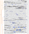
Evoprint of the cis-regulatory DNA used in this study. Bold capital letters represent conservation in either all 12 or 11/12 Drosophila species for which genomic data exists. The Flybase coordinates for the first nucleotide of each row is shown to the right, and the approximate positions of the constructs used is shown to the left (see methods for exact location of these constructs). Transcription factor predicted binding sites within the conserved blocks of DNA were determined using the TESS website (http://www.cbil.upenn.edu/cgi-bin/tess). Blue, Seven-up (Svp) binding sites; gray, Hb binding sites; yellow, Pou domain binding sites, including Nubbin/Pdm2; blue underlines, homeodomain core motifs (ATTA/TAAT) sites.
Fig. 3
Fig. 3
Expression of the endogenous hunchback gene (hb) and enhancer-reporter transgenes (HG series) in neuroblasts and GMCs. Drosophila embryonic NBs first delaminate at early stage 9 and express hb at the time of their delamination; they maintain hb for about an hour until stage 10. In addition, some NBs first delaminate at stage 10 or stage 11, and they also typically express hb for about an hour after delaminating, so that there are scattered Hb+ NBs persisting into stage 11. Nevertheless, the bulk of hb expression occurs at stage 9 and 10. RNA in situ hybridization shown for the indicated genotypes. Black arrows indicate the neuroblast layer in stage 9–11 embryos; white arrowheads indicate the GMC layer in the stage 11 embryos. Only lines HG4-1, HG4-3, and HG4-7 show neuroblast expression (“neuroblast element” label, to right). Scale bar, 20 μm.
Fig. 4
Fig. 4
Expression of the endogenous hunchback gene (hb) and enhancer-reporter transgenes (HG series) in stage 11 neuroblasts. Confocal imaging of embryos stained for GFP (top row) and the neuroblast marker Deadpan (Dpn, bottom row). Lines HG4-1, HG4-3, and HG4-7 show expression in most or all neuroblasts (“neuroblast element” label, at bottom). Two segments are shown, determined by Engrailed staining (not shown). Scale bar, 10 μm.
Fig. 5
Fig. 5
Expression of the endogenous hunchback gene (hb) and enhancer-reporter transgenes (HG series) in neurons of stage 16 embryos, shown in lateral (left) or ventral (right) views. RNA in situ hybridization shown for the indicated genotypes. In the left column, deep neurons (d) are located below the dashed line and superficial neurons (s) are located above the dashed line; examples of deep neurons are shown by black arrows; examples of superficial neurons are shown by white arrowheads; brackets indicate the CNS (right column). Lines HG4-1, HG4-2, HG4-3, HG4-5 and HG4-6 show neuronal expression (“neuron element” label, to right). Scale bar, 20 μm (left column) and 50 μm (right column).
Fig. 6
Fig. 6
Expression of the endogenous hunchback gene (hb) and enhancer-reporter transgenes (HG series) in the stage 16 CNS. Confocal images of the CNS stained for HG reporter construct patterns (GFP, green) and endogenous Hb protein (red). (A–G) Ventral view of deep layer, early-born, Hb+ neurons. Lines HG4-1, HG4-2, HG4-3, HG4-5 and HG4-6 show expression in these Hb+ neurons. (H–N) Ventral view of superficial layer, late-born, Hb-negative neurons. Only line HG4-5 shows strong expression in these Hb-negative neurons; other lines show scattered expression (HG4-1, HG4- 2, HG4-3) or high background due to increasing the confocal gain to try and detect expression (HG4- 4, HG4-6, HG4-7). For A–N, two segments are shown, determined by Engrailed staining in the fourth confocal channel (not shown). (O–Q) Lateral view of a stage 16 CNS showing HG4-5 expression in superficial neurons (O, Q; green GFP staining) and almost no expression in Hb+ deep layer neurons (P, Q; red endogenous Hb staining). Scale bar is 50 μm in A–N, and 10 μm in O–Q.
Fig. 7
Fig. 7
Summary of the cis-regulatory elements controlling hunchback (hb) CNS expression. Grey numbers at top are the Flybase coordinates for the region. Lines shown at right indicate the DNA elements used in the reporter constructs. Color code for the four different regulatory functions discovered in this work are shown in the box, lower left.

References

    1. Brody T, Odenwald WF. Programmed transformations in neuroblast gene expression during Drosophila CNS lineage development. Dev Biol. 2000;226:34–44. - PubMed
    1. Cleary MD, Doe CQ. Regulation of neuroblast competence: multiple temporal identity factors specify distinct neuronal fates within a single early competence window. Genes Dev. 2006;20:429–434. - PMC - PubMed
    1. Doe CQ. Neural stem cells: balancing self-renewal with differentiation. Development. 2008;135:1575–1587. - PubMed
    1. Grosskortenhaus R, Pearson BJ, Marusich A, Doe CQ. Regulation of temporal identity transitions in Drosophila neuroblasts. Dev Cell. 2005;8:193–202. - PubMed
    1. Isshiki T, Pearson B, Holbrook S, Doe CQ. Drosophila neuroblasts sequentially express transcription factors which specify the temporal identity of their neuronal progeny. Cell. 2001;106:511–521. - PubMed

Publication types

MeSH terms

LinkOut - more resources