Review
. 2012 Aug;28(6):1465-75.
doi: 10.1007/s10554-011-9957-4. Epub 2011 Oct 28.

Cardiac magnetic resonance imaging and its electrocardiographs (ECG): tips and tricks

Affiliations
Review

Cardiac magnetic resonance imaging and its electrocardiographs (ECG): tips and tricks

Marcelo Souto Nacif et al. Int J Cardiovasc Imaging. 2012 Aug.

Abstract

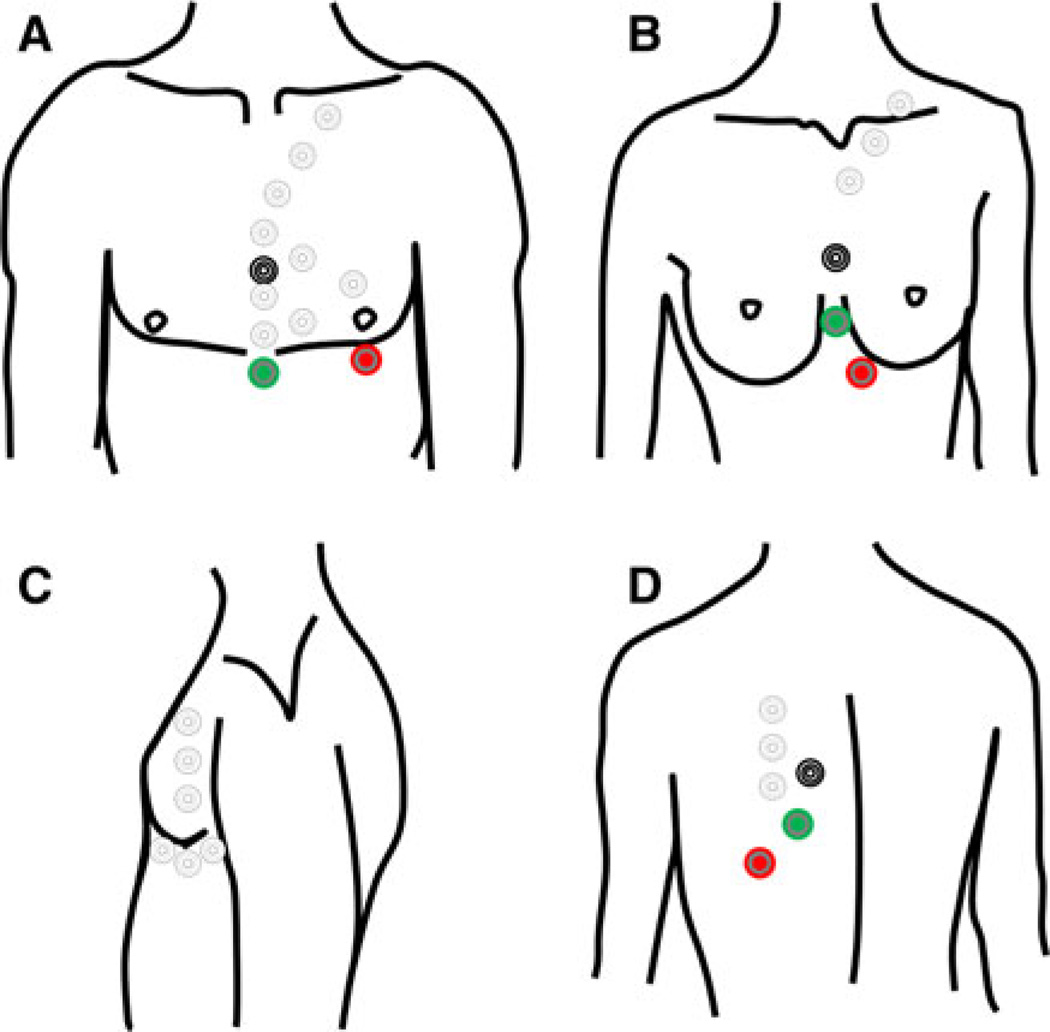
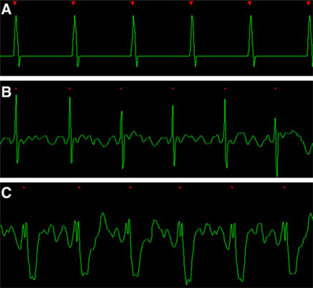
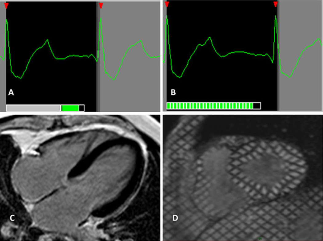
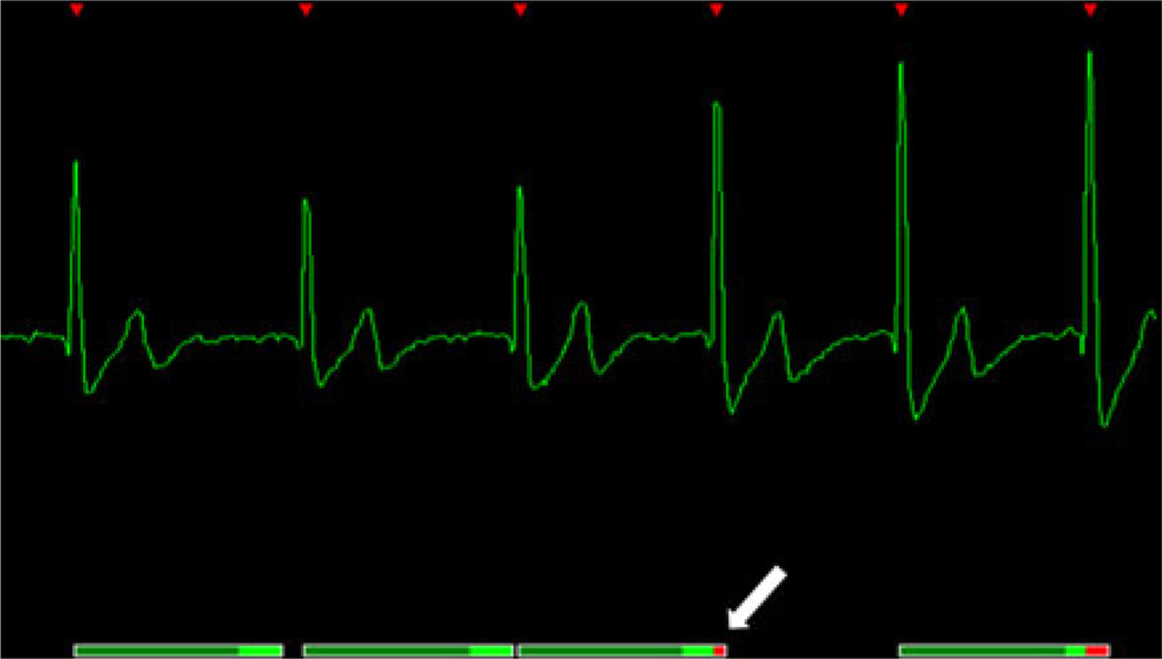
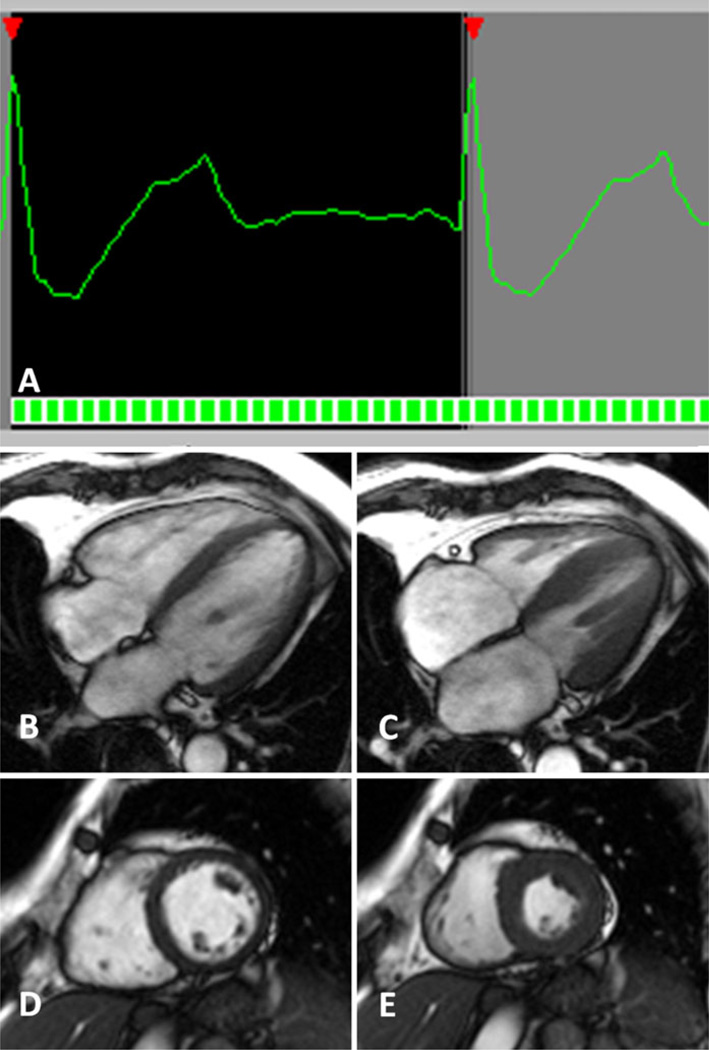
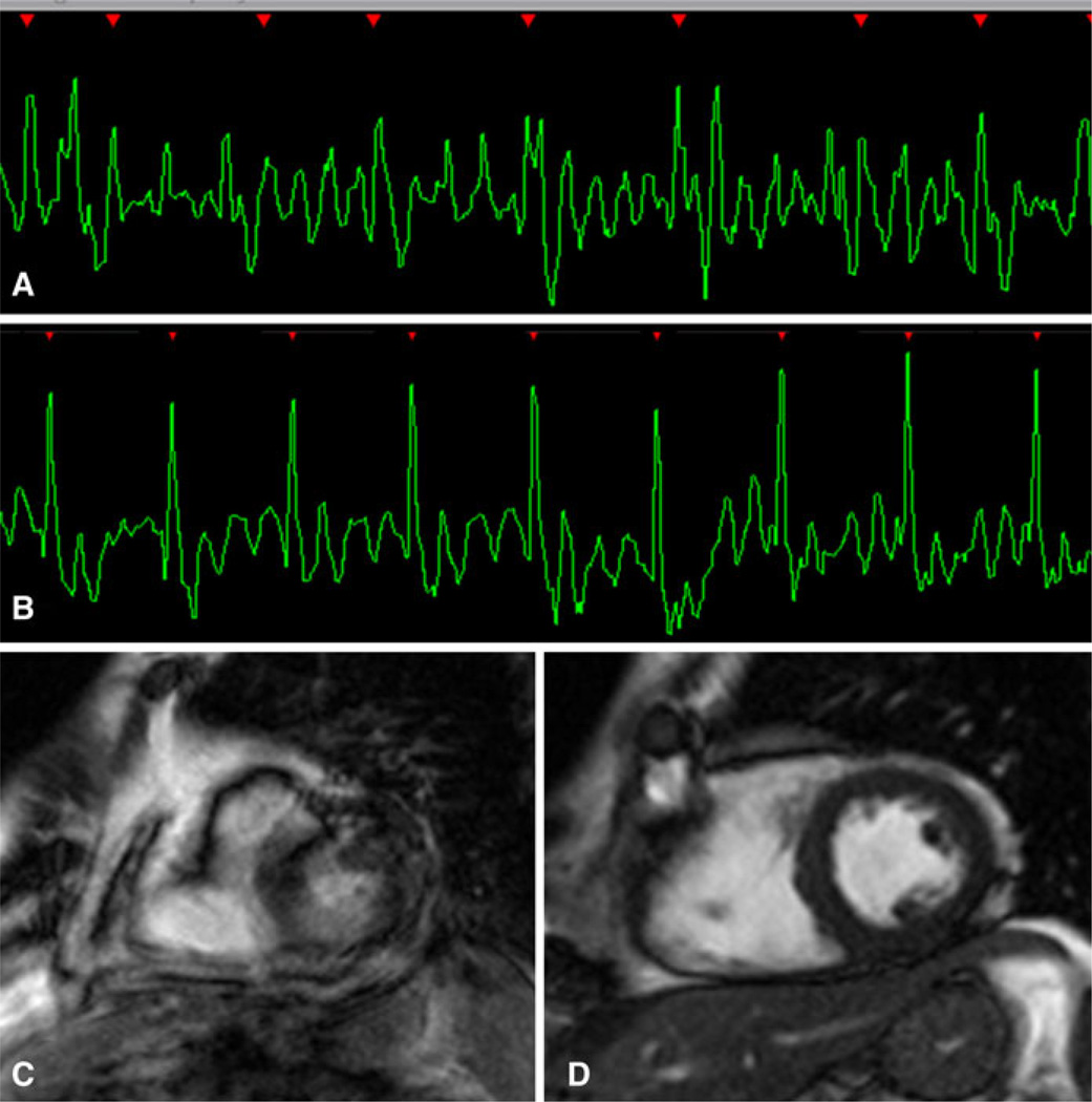
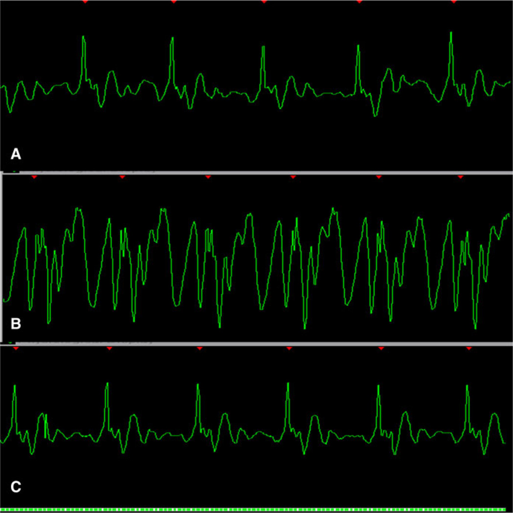
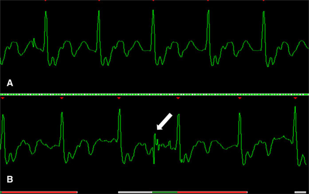
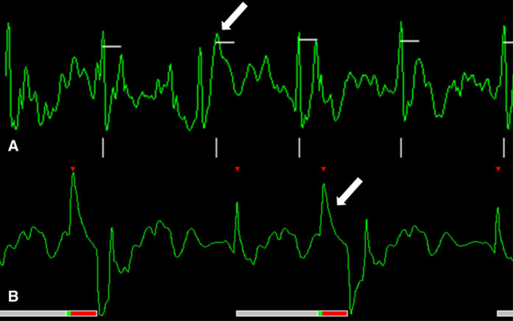
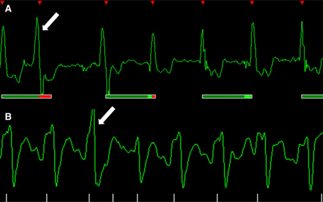
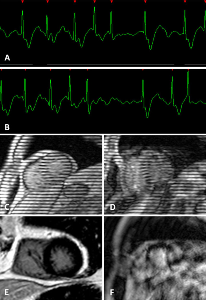
All cardiac magnetic resonance (CMR) techniques aim to create still depictions of a dynamic and ever-adapting organ. Most CMR methods rely on cardiac gating to capture information during fleeting periods of relative cardiac quiescence, at end diastole or end systole, or to acquire partial images throughout the cardiac cycle and average these signals over several heart beats. Since the inception of clinical CMR in the early 1980s, priority has been given to improving methods for image gating. The aim of this work is to provide a basic understanding of the ECG acquisition, demonstrate common ECG-related artifacts and to provide practical methods for overcoming these issues. Meticulous ECG preparation is essential for optimal CMR acquisition and these techniques must be adaptable to the individual patient.


Conflict of interest statement

Conflict of Interest The authors declare that they have no competing interests.

Figures

Fig. 1
Fig. 1
Black, green and red leads denote the standard position of the conventional electrodes. Uncolored circles represent alternative placements which can range from the mid-sternal region to the neck, lateral chest wall and back. a Frontal view. Alignment of the leads along the parasternal midline often increases the R-wave amplitude. b Frontal view. In women, it is often best to avoid the fatty breast tissue, so lead placement can be adapted to breast size and position. c Lateral positions. d Posterior view
Fig. 2
Fig. 2
a An ideal ECG created by a simulation program. The R waves are tall and sensed every time, the R–R intervals are exactly evenly spaced and the intervening baseline is flat without any noise. b A excellent in vivo example. High peak amplitude R-waves are separated by minimal baseline noise. c The QRS complex is inverted; however, the high amplitude of the R-wave will serve as a reliable trigger for the acquisition
Fig. 3
Fig. 3
a Prospective gating for morphological imaging. The signal is acquired during the mid diastolic phase, between the T-wave (cardiac repolarization) and the a-wave (atrial contraction). b Prospective gating for functional imaging. A cine series is acquired beginning with the R-wave. The end of diastole is missed, since the acquisition has to be terminated in order not miss the next R-wave trigger. c Delayed Gadolinium Enhancement is an example of prospective gating. d Tagging cine-MR is an example of prospective cine gating
Fig. 4
Fig. 4
An acquisition window which is placed too close to the next R-wave may reach into the R-wave when the next R–R-interval is only slightly shorter (arrow). This problem particularly occurs during breath hold. Before acquiring the sequence it is necessary to move the acquisition window towards the mid-diastole, usually by shortening the TR time (ms)
Fig. 5
Fig. 5
a Retrospective gating for functional imaging. Complex post-processing methods are used to meld the imaging and ECG signals. The cine series contains all phases of the cardiac cycle. Four chamber cine-MR in end diastole (b) and end systole (c). Short axis cine-MR in end diastole (d) and end systole (e)
Fig. 6
Fig. 6
a Poor electrode placement. Poor electrode placement can result in low amplitude R-wave which is difficult to discern from the baseline noise. These tracings are often mistaken for arrhythmias. Before deciding that a patient’s scan must be cancelled, many electrode positions must be tried (see Fig. 4). b The same patient as in Fig. 5a with a better choice of electrode placement results in a normal ECG trace. c The result of the poor electrode placement on a short axis cine-MR. d The image of the same patient after correction of the ECG placement
Fig. 7
Fig. 7
a A normal tracing is seen in this patient outside the magnet bore. b Once the patient is positioned in the magnet‘s center without changing electrode position a hydrodynamic artifact occurs. c For the patient in Fig. 6a, b, electrode positions on the front and side were unsuccessful in eliminating the ECG artifacts. In this subject the ECG tracing was normalized within the magnet’s bore when the electrodes were place on the back
Fig. 8
Fig. 8
Depending on the intensity and how the sequence changes the magnet field with the radiofrequency pulse we can see that in the same patient the artifacts can occur. a Cine-MR retrospective gating without artifact. b Spectroscopy CMR prospective gating using two R–R intervals with a base line noise correlated to the pulse sequence (white arrow). If the amplitude of the R wave is not sufficient it can lead to a false triggering (not in this case)
Fig. 9
Fig. 9
Two different cases. a A low amplitude R wave is skipped (white arrow) by the scanner when the sequence pulse is done adding noise to a T wave that is wrongly triggered. b A low amplitude R wave is skipped by the scanner as in figure (a), however now triggering on the A wave
Fig. 10
Fig. 10
Extra heartbeat. a Observe the difference within the second (white arrow) and third QRS complex. b Premature ventricular contraction (white arrow) observed in the third QRS complex. When extra-beats occur the scanner does not trigger correctly and the image appears blurred depending on the frequency of this occurrence
Fig. 11
Fig. 11
Atrial fibrillation. a, b Length of the R–R intervals vary to an extent that proper k-space sampling is hampered. Cine-MR tagging with a c normal ECG trace and on an d atrial fibrillation patient. Delayed enhancement with a e normal ECG trace and on an f atrial fibrillation patient

References

    1. Lanzer P, Botvinick EH, Schiller NB, et al. Cardiac imaging using gated magnetic resonance. Radiology. 1984;150:121–127. - PubMed
    1. Amoore JN, Ridgway JP. A system for cardiac and respiratory gating of a magnetic resonance imager. Clin Phys Physiol Meas. 1989;10:283–286. - PubMed
    1. McNamara MT, Higgins CB. Cardiovascular applications of magnetic resonance imaging. Magn Reson Imaging. 1984;2:167–183. - PubMed
    1. Osbakken M, Yuschok T. Evaluation of ventricular function with gated cardiac magnetic resonance imaging. Cathet Cardiovasc Diagn. 1986;12:156–160. - PubMed
    1. Stark DD, Higgins CB, Lanzer P, et al. Magnetic resonance imaging of the pericardium: normal and pathologic findings. Radiology. 1984;150:469–474. - PubMed

Publication types