. 2012 Jan;123(1):105-17.
doi: 10.1007/s00401-011-0893-0. Epub 2011 Oct 28.

Dual modification of Alzheimer's disease PHF-tau protein by lysine methylation and ubiquitylation: a mass spectrometry approach

Affiliations

Dual modification of Alzheimer's disease PHF-tau protein by lysine methylation and ubiquitylation: a mass spectrometry approach

Stefani N Thomas et al. Acta Neuropathol. 2012 Jan.

Abstract

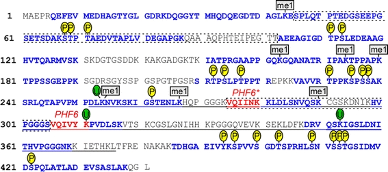
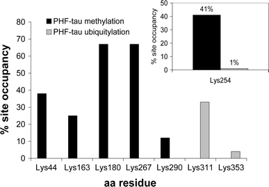
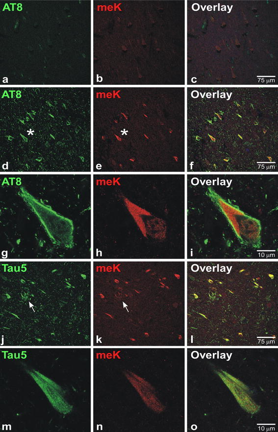
In sporadic Alzheimer's disease (AD), neurofibrillary lesion formation is preceded by extensive post-translational modification of the microtubule associated protein tau. To identify the modification signature associated with tau lesion formation at single amino acid resolution, immunopurified paired helical filaments were isolated from AD brain and subjected to nanoflow liquid chromatography-tandem mass spectrometry analysis. The resulting spectra identified monomethylation of lysine residues as a new tau modification. The methyl-lysine was distributed among seven residues located in the projection and microtubule binding repeat regions of tau protein, with one site, K254, being a substrate for a competing lysine modification, ubiquitylation. To characterize methyl lysine content in intact tissue, hippocampal sections prepared from post mortem late-stage AD cases were subjected to double-label confocal fluorescence microscopy using anti-tau and anti-methyl lysine antibodies. Anti-methyl lysine immunoreactivity colocalized with 78 ± 13% of neurofibrillary tangles in these specimens. Together these data provide the first evidence that tau in neurofibrillary lesions is post-translationally modified by lysine methylation.


Figures

Fig. 1
Fig. 1
Summary of modification sites identified by LC–MS/MS on immunopurified PHF-tau. The sequence shown is that of human 2N4R tau (NCBI accession number NP_005901). Bold sequence coverage; Red PHF6 and PHF6* motifs; dashed lines segments encoded by alternatively spliced exons 2, 3, and 10; underline repeat region (as defined in [23]); P Phosphorylated sites, U Ubiquitylated sites, me1 monomethylated sites identified from MS analysis reported herein and in [13]
Fig. 2
Fig. 2
Relative abundance of PHF-tau methylation and ubiquitylation. Relative abundance was calculated based on the spectral counts of the modified peptide/(modified peptide + unmodified peptide). Only sites having total spectral counts >3 are shown. Inset quantifies K254 methylation (41%) and ubiquitylation (1%). K254 was the most abundant methylated site identified on PHF-tau (spectral count = 17) and the only site on PHF-tau identified in methylated and ubiquitylated forms
Fig. 3
Fig. 3
MS/MS characterization of K254 modification site. Spectra for peptide aa241–254 derived from Lys-C cleavage of PHF-tau identified in (a) unmodified and (b) methylated (asterisks site of methylation) forms. c Spectrum for tryptic peptide aa243–257 identified in ubiquitylated form (carets site of ubiquitylation). Trypsin did not cleave ubiquitylated aa243–257 at K254 owing to its conjugation to the C-terminal di-Gly fragment of ubiquitin
Fig. 4
Fig. 4
MS/MS identification of S262 phosphorylated and K267 methylated PHF-tau peptides. Spectra for PHF-tau derived peptide aa258–267 identified in a unmodified, b monomethylated (asterisks site of methylation), c Ser phosphorylated (at sign site of phosphorylation), and d doubly modified (asterisks site of methylation; at sign site of phosphorylation) forms
Fig. 5
Fig. 5
Anti-meK antibody specificity. Aliquots of unmodified (−) and in vitro methylated (+) 2N4R tau were separated by SDS-PAGE (8% polyacrylamide) and either a stained with Coomassie Blue (500 ng tau proteins), or b subjected to immunoblot analysis with anti-meK antibody (100 ng tau protein). Molecular mass calibration markers (M) are shown in units of kDa. High-stoichiometry reductive methylation reduced recombinant 2N4R migration on SDS-PAGE. To further test specificity, antibodies AT8 (c, f; green channel) and anti-meK (d, g; red channel) were pre-adsorbed with either unmodified (c–e) or in vitro methylated (f–h) 2N4R tau, and then subjected to double-label confocal immunofluorescence microscopy on sections of AD hippocampus. Lesions labeled with both AT8 and anti-meK are marked by asterisks. AT8 labeling was unaffected by preadsorption with either unmodified or methylated tau, whereas anti-meK immunoreactivity was diminished by pre-adsorption with methylated (f–h; carets), but not unmodified tau (c–e)
Fig. 6
Fig. 6
Anti-meK immunoreactivity colocalizes with neurofibrillary lesions in AD hippocampus. Double-label confocal images of hippocampal sections (CA1 region) stained with anti-tau mouse monoclonal antibodies AT8 (a–i; green channel) or Tau5 (j–o; green channel) along with the rabbit anti-meK antibody (red channel). In AD cases, low magnification images (40× objective,1× zoom) show colocalization of anti-meK immunoreactivity with NFTs (d–f; asterisks) and a neuritic plaque (j–l; arrows). High magnification images (100× objective, 3× zoom) show typical morphology of NFTs (g–i and m–o). Image overlays highlight pixel overlap between anti-meK and AT8 (d–i) and Tau5 (j–o) immunoreactivity. Anti-meK immunoreactivity colocalized extensively with NFTs in all fields examined. In contrast, AT8 and anti-meK immunoreactivity did not colocalize in hippocampal sections prepared from cognitively normal cases (a–c)

References

    1. Alonso A, Zaidi T, Novak M, Grundke-Iqbal I, Iqbal K. Hyperphosphorylation induces self-assembly of tau into tangles of paired helical filaments/straight filaments. Proc Natl Acad Sci USA. 2001;98:6923–6928. doi: 10.1073/pnas.121119298. - DOI - PMC - PubMed
    1. Augustinack JC, Schneider A, Mandelkow EM, Hyman BT. Specific tau phosphorylation sites correlate with severity of neuronal cytopathology in Alzheimer’s disease. Acta Neuropathol (Berl) 2002;103:26–35. doi: 10.1007/s004010100423. - DOI - PubMed
    1. Beausoleil SA, Jedrychowski M, Schwartz D, et al. Large-scale characterization of HeLa cell nuclear phosphoproteins. Proc Natl Acad Sci USA. 2004;101:12130–12135. doi: 10.1073/pnas.0404720101. - DOI - PMC - PubMed
    1. Biernat J, Gustke N, Drewes G, Mandelkow EM, Mandelkow E. Phosphorylation of Ser262 strongly reduces binding of tau to microtubules: distinction between PHF-like immunoreactivity and microtubule binding. Neuron. 1993;11:153–163. doi: 10.1016/0896-6273(93)90279-Z. - DOI - PubMed
    1. Braak H, Alafuzoff I, Arzberger T, Kretzschmar H, Del Tredici K. Staging of Alzheimer disease-associated neurofibrillary pathology using paraffin sections and immunocytochemistry. Acta Neuropathol. 2006;112:389–404. doi: 10.1007/s00401-006-0127-z. - DOI - PMC - PubMed

Publication types

MeSH terms

LinkOut - more resources