. 2011 Dec 9;286(49):42099-42104.
doi: 10.1074/jbc.C111.294462. Epub 2011 Oct 27.

Crystal structure of the chromodomain helicase DNA-binding protein 1 (Chd1) DNA-binding domain in complex with DNA

Affiliations

Crystal structure of the chromodomain helicase DNA-binding protein 1 (Chd1) DNA-binding domain in complex with DNA

Amit Sharma et al. J Biol Chem. .

Abstract

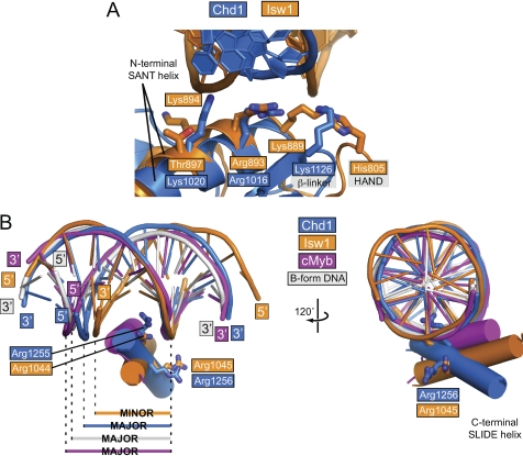
Chromatin remodelers are ATP-dependent machines that dynamically alter the chromatin packaging of eukaryotic genomes by assembling, sliding, and displacing nucleosomes. The Chd1 chromatin remodeler possesses a C-terminal DNA-binding domain that is required for efficient nucleosome sliding and believed to be essential for sensing the length of DNA flanking the nucleosome core. The structure of the Chd1 DNA-binding domain was recently shown to consist of a SANT and SLIDE domain, analogous to the DNA-binding domain of the ISWI family, yet the details of how Chd1 recognized DNA were not known. Here we present the crystal structure of the Saccharomyces cerevisiae Chd1 DNA-binding domain in complex with a DNA duplex. The bound DNA duplex is straight, consistent with the preference exhibited by the Chd1 DNA-binding domain for extranucleosomal DNA. Comparison of this structure with the recently solved ISW1a DNA-binding domain bound to DNA reveals that DNA lays across each protein at a distinct angle, yet contacts similar surfaces on the SANT and SLIDE domains. In contrast to the minor groove binding seen for Isw1 and predicted for Chd1, the SLIDE domain of the Chd1 DNA-binding domain contacts the DNA major groove. The majority of direct contacts with the phosphate backbone occur only on one DNA strand, suggesting that Chd1 may not strongly discriminate between major and minor grooves.


Figures

FIGURE 1.
FIGURE 1.
Overview of the Chd1 DNA-binding domain bound to DNA and comparison with the Isw1-DNA structure. A, backbone (left) and graphic (right) representations of the S. cerevisiae Chd1 DNA-binding domain bound to a dodecameric DNA duplex. Four residues that make up part of the DNA-binding surface are highlighted as magenta spheres (shown also in Figs. 2 and 3). In the graphic, the dotted lines represent the expected position of the DNA backbone continued in a straight trajectory from the duplex observed in the crystal. Residues on the HL1 extension unique to Chd1 that appear positioned to contact DNA are shown as yellow sticks. The orange circles on the DNA graphic indicate common points of contact from the SANT and SLIDE domains of Chd1 and Isw1. B, backbone (left) and graphic (right) representations for the S. cerevisiae Isw1 DNA-binding domain in complex with a DNA duplex (PDB code 2Y9Z (16)), shown in an orientation that aligns the Isw1 SLIDE domain with that of Chd1 in A. Four residues that appear to be structurally analogous to those highlighted for Chd1 in A are shown as magenta spheres. Note that the Ioc3p subunit of the ISW1a complex, which is bound beneath the HAND-SANT-SLIDE domain in this orientation, is not shown for clarity.
FIGURE 2.
FIGURE 2.
Similar residues from the Chd1 and Isw1 SANT-SLIDE domains contact the phosphate backbone of DNA. A, alignment of the SANT domain from Chd1 (blue) and Isw1 (orange, PDB code 2Y9Z (16)) shows that although the DNA crosses each domain differently, similarly positioned residues on the N-terminal SANT helix contact the phosphate backbone. Lys-1126 on the β-linker, which is unique to Chd1 proteins, appears to take the place of Lys-889, a conserved basic position in ISWI-type remodelers on the first turn of the N-terminal SANT helix (11). B, alignment of DNA duplexes reveals that the C-terminal SLIDE helix of Chd1 falls in the DNA major groove, in contrast to the minor groove binding observed for Isw1 (16). For this alignment, the DNA strand making direct contact to the Chd1 SLIDE domain was used as a reference. Also shown is the C-terminal helix of the c-Myb R2 domain bound to a DNA duplex (magenta, PDB code 1H89 (14)) and an idealized B-form DNA duplex (gray). Note that the duplexes bound to Chd1 (blue) and Isw1 (orange) have the opposite polarity, which reflects the position of the C-terminal SLIDE helix against the DNA major or minor grooves, respectively.
FIGURE 3.
FIGURE 3.
The Chd1 SLIDE domain primarily makes direct contacts with only one of the two DNA strands. A, direct contacts to the DNA phosphate backbone of one strand are made with residues from the C-terminal SLIDE helix and a loop following the spacer helix. Water molecules are shown as red spheres, hydrogen bonds are shown as green dashes, and hydrophobic contacts are shown as yellow dots. B, water-mediated contacts with DNA on the other side of the C-terminal SLIDE helix. Also shown is a water-mediated contact from a tyrosine on the SANT domain (green) and direct contacts from the β-linker (yellow) and spacer helix (red). Electron density maps of the protein-DNA complex for the views shown in A and B are available as supplemental Movies S1 and S2.

References

    1. Gangaraju V. K., Bartholomew B. (2007) Mutat. Res. 618, 3–17 - PMC - PubMed
    1. Ryan D. P., Owen-Hughes T. (2011) Curr. Opin. Chem. Biol. 15, 649–656 - PMC - PubMed
    1. Delmas V., Stokes D. G., Perry R. P. (1993) Proc. Natl. Acad. Sci. U.S.A. 90, 2414–2418 - PMC - PubMed
    1. Marfella C. G., Imbalzano A. N. (2007) Mutat. Res. 618, 30–40 - PMC - PubMed
    1. Walfridsson J., Khorosjutina O., Matikainen P., Gustafsson C. M., Ekwall K. (2007) EMBO J. 26, 2868–2879 - PMC - PubMed

Publication types

MeSH terms

Associated data