

JCV agnoprotein-induced reduction in CXCL5/LIX secretion by oligodendrocytes is associated with activation of apoptotic signaling in neurons
- PMID: 22034072
- PMCID: PMC3296900
- DOI: 10.1002/jcp.23065
JCV agnoprotein-induced reduction in CXCL5/LIX secretion by oligodendrocytes is associated with activation of apoptotic signaling in neurons
Abstract
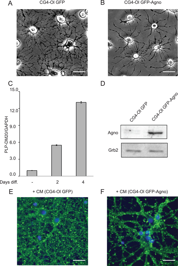
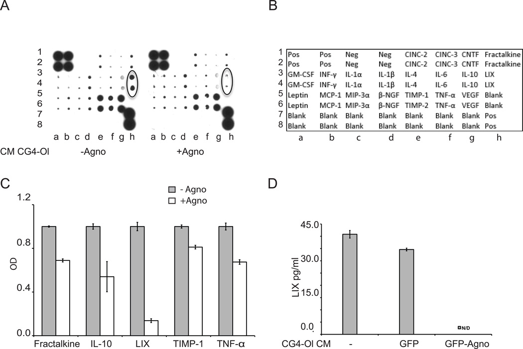
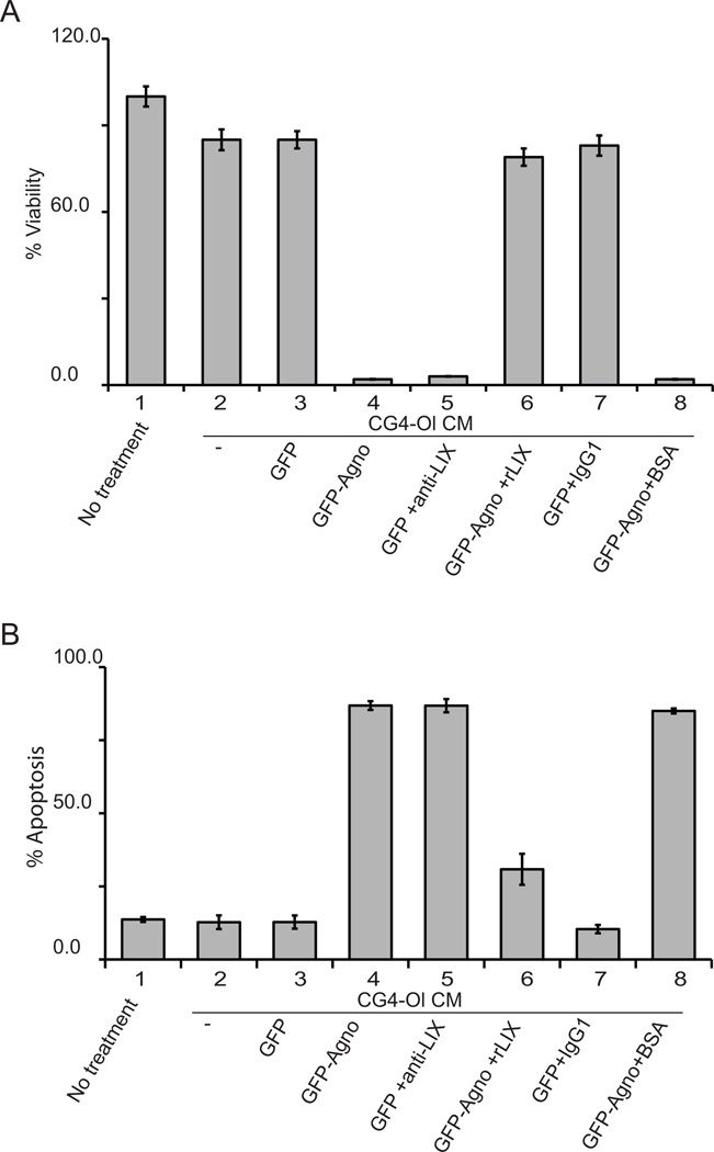
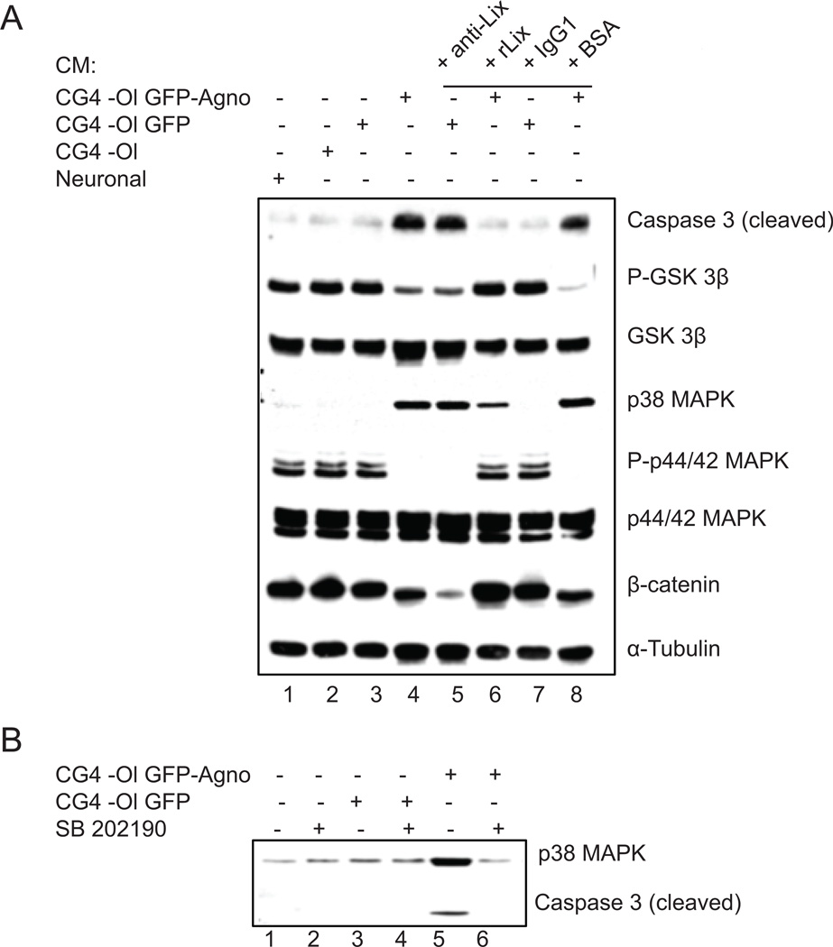
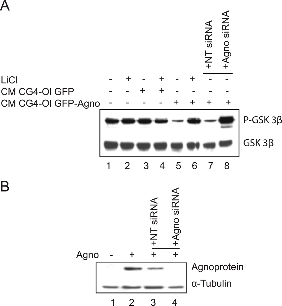
An indispensable role for oligodendrocytes in the protection of axon function and promotion of neuronal survival is strongly supported by the finding of progressive neuron/axon degeneration in human neurological diseases that affect oligodendrocytes. Imaging and pathological studies of the CNS have shown the presence of neuroaxonal injury in progressive multifocal leukoencephalopathy (PML), a demyelinating disease of the CNS, resulting from destruction of oligodendrocytes upon productive replication of the pathogenic neurotropic polyomavirus JC. Here, we examined the extracellular factors involved in communication between oligodendrocytes and neurons. Culturing cortical neurons with conditioned medium (CM) from rat CG4 oligodendrocytic cells that express the JCV agnoprotein showed that CXCL5/LIX, which is a chemokine closely related to the human CXCL5/ENA78 and CXCL6/GCP-2 chemokines, is essential for neuronal cell survival. We found that in CM from agnoprotein-producing CG-4 cells level of CXC5/LIX is decreased compared to control cells. We also demonstrated that a reduced expression of CXCL5/LIX by CG4 GFP-Agno cells triggered a cascade of signaling events in cortical neurons. Analysis of mitogen-activated protein kinases (MAPK) and glycogen synthase kinase (GSK3) pathways showed that they are involved in mechanisms of neuronal apoptosis in response to the depletion of CXCL5/LIX signaling. These data suggest that agnoprotein-induced dysregulation of chemokine production by oligodendrocytes may contribute to neuronal/axonal injury in the pathogenesis of PML lesions.
Copyright © 2011 Wiley Periodicals, Inc.
Figures
Similar articles
-
Expression of JC virus agnoprotein in progressive multifocal leukoencephalopathy brain.Acta Neuropathol. 2002 Aug;104(2):130-6. doi: 10.1007/s00401-002-0526-8. Epub 2002 May 24. Acta Neuropathol. 2002. PMID: 12111355
-
JC virus agnoprotein inhibits in vitro differentiation of oligodendrocytes and promotes apoptosis.J Virol. 2008 Feb;82(3):1558-69. doi: 10.1128/JVI.01680-07. Epub 2007 Nov 7. J Virol. 2008. PMID: 17989177 Free PMC article.
-
The human polyoma JC virus agnoprotein acts as a viroporin.PLoS Pathog. 2010 Mar 12;6(3):e1000801. doi: 10.1371/journal.ppat.1000801. PLoS Pathog. 2010. PMID: 20300659 Free PMC article.
-
Potential mechanisms of the human polyomavirus JC in neural oncogenesis.J Neuropathol Exp Neurol. 2008 Aug;67(8):729-40. doi: 10.1097/NEN.0b013e318180e631. J Neuropathol Exp Neurol. 2008. PMID: 18648329 Free PMC article. Review.
-
[Progressive multifocal leukoencephalopathy. Demyelinating viral disease--common complication of AIDS].Lakartidningen. 2001 Sep 26;98(39):4206-11, 4213. Lakartidningen. 2001. PMID: 11680156 Review. Swedish.
Cited by
-
Agnoprotein of polyomavirus BK interacts with proliferating cell nuclear antigen and inhibits DNA replication.Virol J. 2015 Feb 1;12:7. doi: 10.1186/s12985-014-0220-1. Virol J. 2015. PMID: 25638270 Free PMC article.
-
Loss of APP in mice increases thigmotaxis and is associated with elevated brain expression of IL-13 and IP-10/CXCL10.Physiol Behav. 2021 Oct 15;240:113533. doi: 10.1016/j.physbeh.2021.113533. Epub 2021 Jul 19. Physiol Behav. 2021. PMID: 34293404 Free PMC article.
-
Mitochondrial Impairment in Oligodendroglial Cells Induces Cytokine Expression and Signaling.J Mol Neurosci. 2019 Feb;67(2):265-275. doi: 10.1007/s12031-018-1236-6. Epub 2018 Dec 13. J Mol Neurosci. 2019. PMID: 30547416
-
CC and CXC chemokines play key roles in the development of polyomaviruses related pathological conditions.Virol J. 2021 Jun 3;18(1):111. doi: 10.1186/s12985-021-01582-4. Virol J. 2021. PMID: 34082771 Free PMC article. Review.
-
Nuclear magnetic resonance structure revealed that the human polyomavirus JC virus agnoprotein contains an α-helix encompassing the Leu/Ile/Phe-rich domain.J Virol. 2014 Jun;88(12):6556-75. doi: 10.1128/JVI.00146-14. Epub 2014 Mar 26. J Virol. 2014. PMID: 24672035 Free PMC article.
References
-
- Atta UR, Harvey K, Siddiqui RA. Interleukin-8: an autocrine inflammatory mediator. Curr Pharm Des. 1999;5:241–253. - PubMed
-
- Bajetto A, Bonavia R, Barbero S, Schettini G. Characterization of chemokines and their receptors in the central nervous system: physiopathological implications. J. Neurochem. 2002;82:1311–1329. - PubMed
-
- Berger JR, Kaszovitz B, Post MJ, Dickinson G. Progressive multifocal leukoencephalopathy associated with human immunodeficiency virus infection. A review of the literature with a report of sixteen cases. Ann. Intern Med. 1987;107:78–87. - PubMed
-
- Berger JR, Houff S. Progressive multifocal leukoencephalopathy: lessons from AIDS and natalizumab. Neurol Res. 2006;28:299–305. - PubMed
-
- Berger JR. Progressive multifocal leukoencephalopathy. Curr Neurol Neurosci Rep. 2007;7:461–469. - PubMed
Publication types
MeSH terms
Substances
Grants and funding
LinkOut - more resources
Full Text Sources
